
|
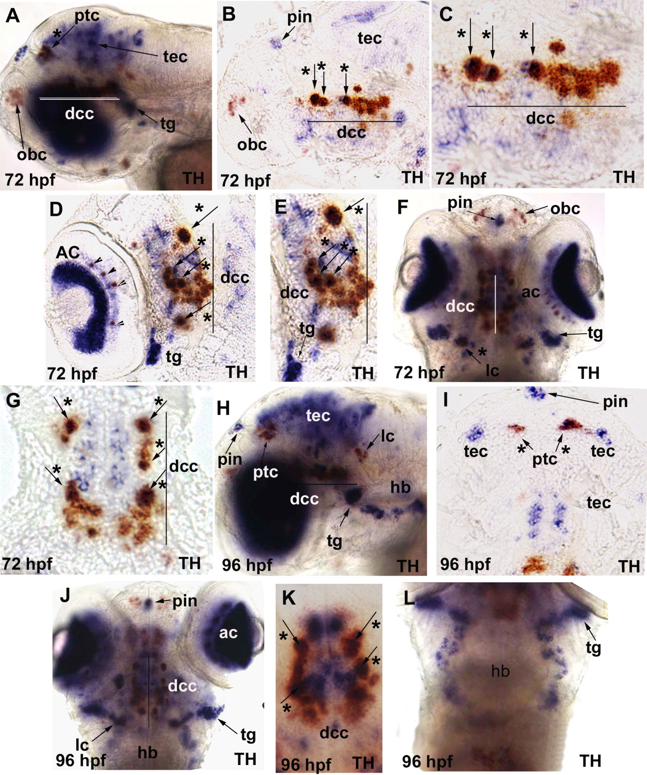
Fig. 4 In situ hybridization analysis of chrna6 RNA expression in 72 hours postfertilization (hpf) and 96 hpf zebrafish embryos. The purple stain represents chrna6 neuronal nicotinic acetylcholine receptor (nAChR) subunit mRNA in all panels, and the orange labeling is tyrosine hydroxylase (TH) mRNA. All images are lateral views with the anterior to the left unless otherwise noted. Arrows point to specific brain regions, arrowheads point to amacrine cells in the retina, and arrows with an asterisk denote co-labeling with TH. Line denotes diencephalic catecholaminergic cluster in A-G. A: At 72 hpf, chrna6 was now localized heavily to the eye and tectum, pineal, trigeminal ganglion, and colocalization with TH in the diencephalic catecholaminergic cluster and pretectal catecholaminergic cluster. B: At 72 hpf, midsagittal section, showed chrna6 expression in pineal, tectum, and colocalization with TH in a subset of cells in the diencephalic catecholaminergic cluster, and no expression in the olfactory bulb catecholaminergic cluster. C: At 72 hpf, midsagittal section, higher magnification of B, colocalization of chrna6 with TH in the diencephalic catecholaminergic cluster. D: At 72 hpf, sagittal section, chrna6 expression in amacrine cells of the retina, retinal ganglion cells, trigeminal ganglion, and colocalization with TH in a subset of cells in the diencephalic catecholaminergic cluster. E: At 72 hpf, sagittal section higher magnification of D, colocalization of chrna6 with TH in the diencephalic catecholaminergic cluster and expression in trigeminal ganglion. F: At 72 hpf, dorsal view of whole animal, showed chrna6 expression in retina, trigeminal ganglion, pineal, and colocalization with TH in the diencephalic catecholaminergic cluster and locus coeruleus. G: At 72 hpf, longitudinal section, showed chrna6 expression in a subset of TH+ cells in the diencephalic catecholaminergic cluster. H: At 96 hpf, expression was evident in retina, pineal, tectum, trigeminal ganglion, a subset of cells in the hindbrain consistent with cranial sensory neurons, and colocalization with TH in the diencephalic catecholaminergic cluster, locus coeruleus, and pretectal catecholaminergic cluster. I: At 96 hpf, longitudinal section, demonstrated colocalization of TH and chrna6 expression in the pretectal area, with pineal and tectum labeled. J: At 96 hpf, dorsal view whole-mount, chrna6 expression shown in retina, pineal, trigeminal ganglion, diencephalic catecholaminergic cluster, and locus coeruleus. K: At 96 hpf, dorsal view whole embryo, chrna6 colocalization with TH in the diencephalic catecholaminergic cluster. L: At 96 hpf, dorsal view whole embryo, chrna6 expression in hindbrain neurons consistent with the localization of cranial sensory neurons. ac, amacrine cells in the retina; dcc, diencephalic catecholaminergic cluster; hb, hindbrain nuclei; lc, locus coereuleus; obc, olfactory bulb catecholaminergic cluster; ptc, pretectal catecholaminergic cluster; pin, pineal; tec, tectum; tg, trigeminal ganglion.