
|
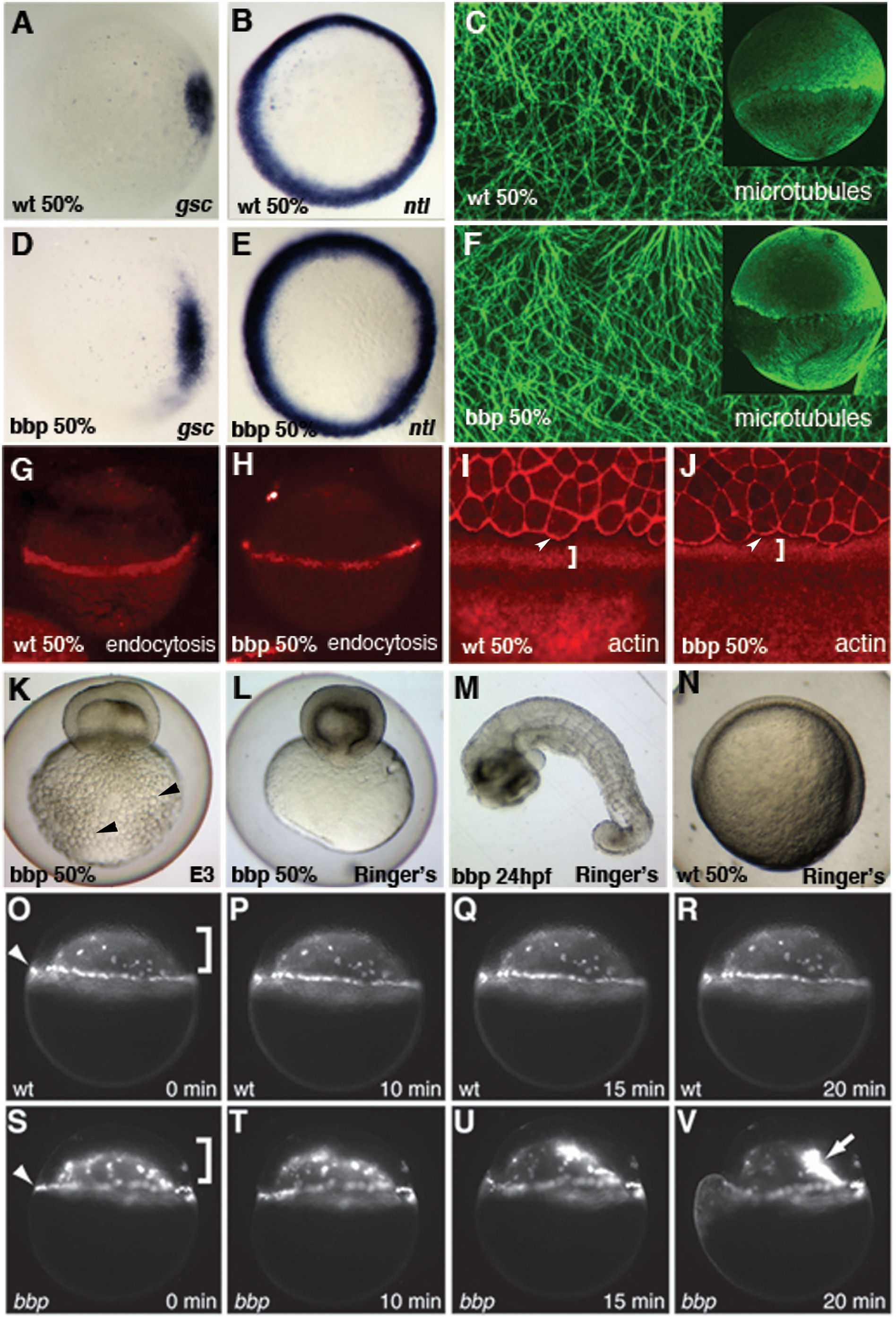
Fig. 3 Characterization of bbp phenotype.
In situ hybridization of goosecoid (gsc) in WT (A) and bbp (D) and no tail (ntl) in WT (B) and bbp (E) embryos at 50% epiboly. Microtubule array forms normally in bbp embryo (F) at the approach to 50% epiboly as in WT (C). G, H Internalization of Rhodamine-dextran through endocytosis in the YSL at 50% epiboly in WT (G) and a bbp (H) embryo. Rhodamine-Phalloidin staining in WT (I) and bbp (J) embryos at 50% epiboly. White brackets show punctate actin band in YSL, arrowheads indicate actin at EVL margin. Hypertonic Ringer's solution extended development of constricting bbp embryos. bbp embryo ni E3 medium constricts at 50% epiboly and yolk globules (black arrowheads) burst through the yolk membrane (K). bbp embryo reared in hypertonic medium constricts, while maintaining yolk membrane integrity (L), allowing the blastoderm to heal and survive to 24 hpf (M). Mutants can undergo gastrulation movements and generate a general body plan at 24 hpf. WT sibling reared in hypertonic medium develops normally, shown at 50% epiboly (N). YSN of WT embryo labeled with Sytox Green (O–R); both I-YSN (bracket) and E-YSN (arrowhead) change little during this short time interval. (S–V) bbp I-YSN are initially distributed normally under the blastoderm, but retract rapidly to a small region of the I-YSL (arrow) when the margin constricts, just before yolk cell rupture.