
|
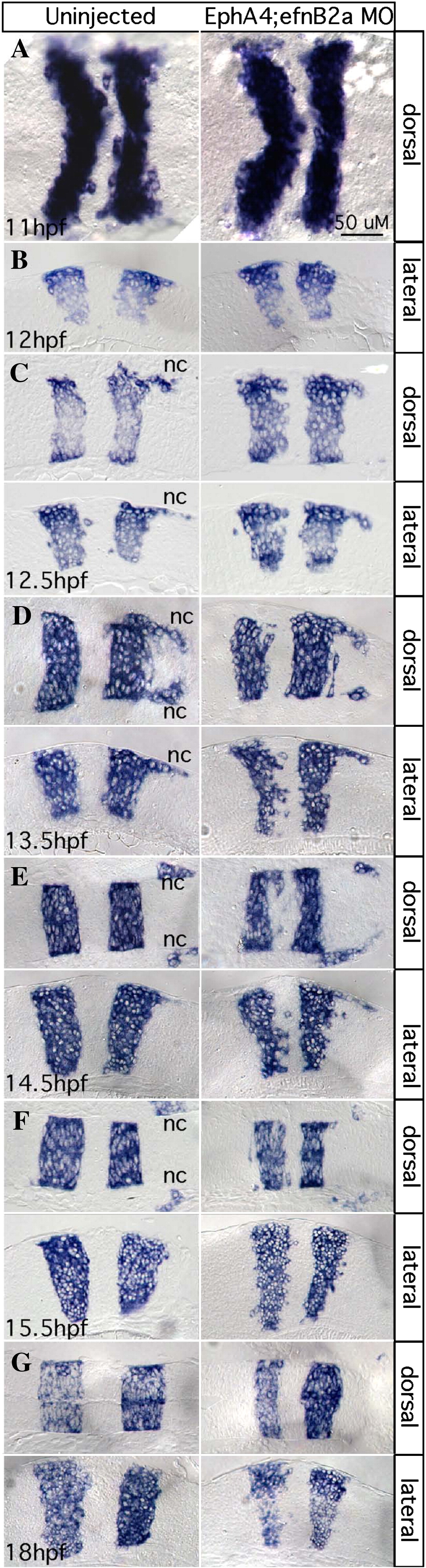
Fig. 3 EphA4 and EfnB2a mediate hindbrain boundary sharpening during the neural keel stage. Whole mount (A) and 10 μm plastic sections (B–G) of RNA in situ hybridizations with a krox20/egr2b riboprobe. Anterior is to the left, dorsal or lateral views are indicated. Left panels show uninjected control embryos; right panels show stage-matched EphA4; EfnB2a double MO embryos. (A) Neural plate stage (N = 20); (B) neural plate to neural keel transition; (C,D) successive neural keel stages; (E,F) successive neural rod stages; (G) neural tube stage. Nc: r5-derived neural crest. Representative sections for each timepoint were chosen from at least 8 identically treated embryos. Scale bar: 50 μm.
Reprinted from Developmental Biology, 327(2), Kemp, H.A., Cooke, J.E., and Moens, C.B., EphA4 and EfnB2a maintain rhombomere coherence by independently regulating intercalation of progenitor cells in the zebrafish neural keel, 313-326, Copyright (2009) with permission from Elsevier. Full text @ Dev. Biol.