
Fig. 1
|
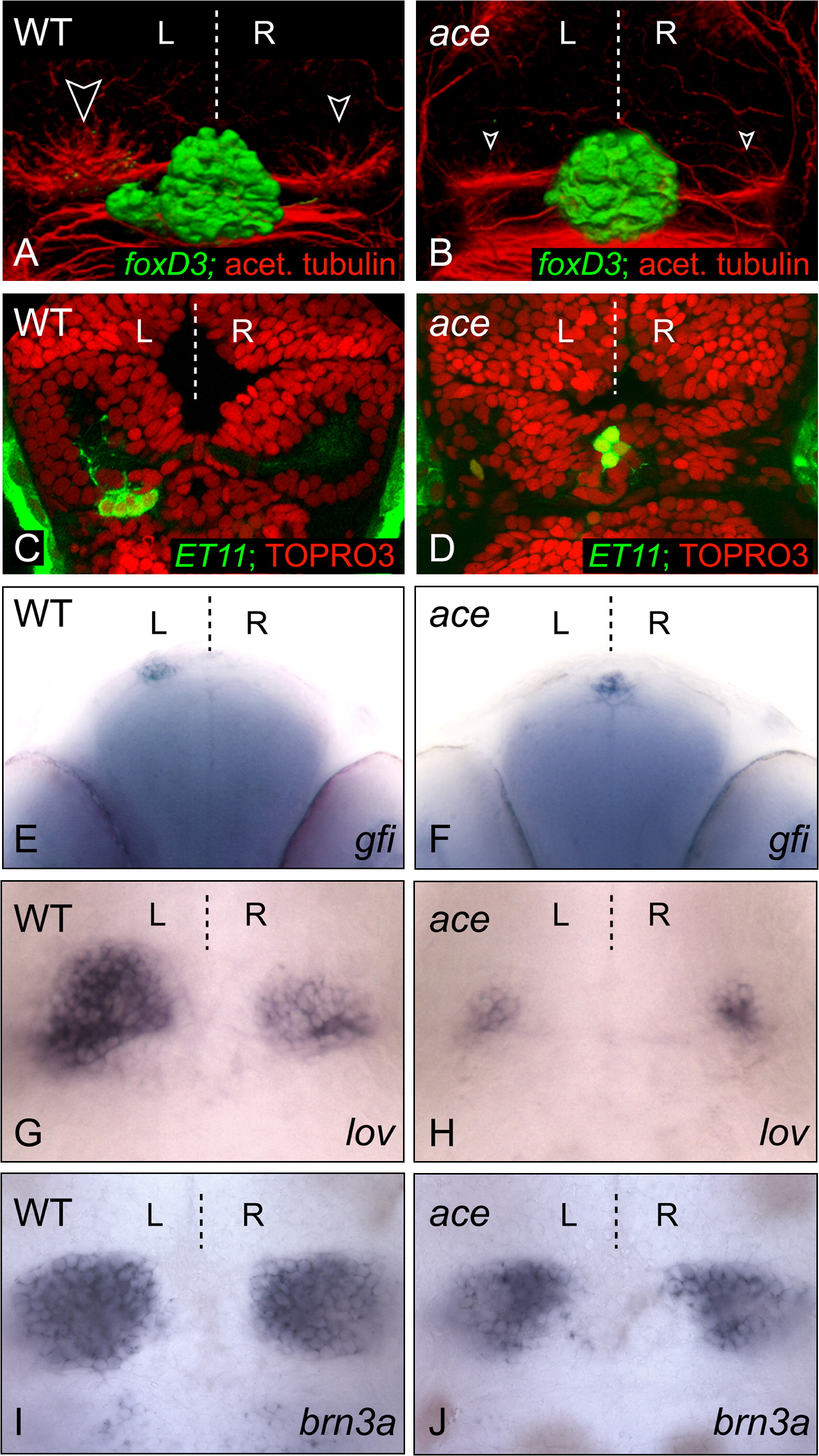
Fig. 1 The fgf8 Mutant Has a Symmetric Epithalamus
(A–D) Dorsal views of confocal images of the epithalamus in wild-type and ace 3 dpf embryos, with anterior to the top. (A and B) 3D reconstructions of pineal/parapineal nuclei and axons [green, Tg(foxD3:GFP)] and neuropil of the habenular nuclei (red, anti-acetylated tubulin; white-edged arrowheads). (C and D) The parapineal-specific marker Tg(ET11:GFP) is present (green), but expressing cells remain at the midline in the ace embryo compared with those of the wild-type embryo at 3 dpf. Brain morphology is visualized using the nuclear marker TOPRO3 (red). A single z-slice is presented for each example. (E and F) Frontal view of parapineal-specific gfi expression in wild-type and ace 3 dpf embryos, with dorsal to the top. Parapineal cells are at the midline in ace (F). (G–J) Dorsal views of habenular lov (G and H) and brn3a (I and J) expression in wild-type and ace embryos at 4 dpf; expression of both markers is reduced in left and right habenulae.