
|
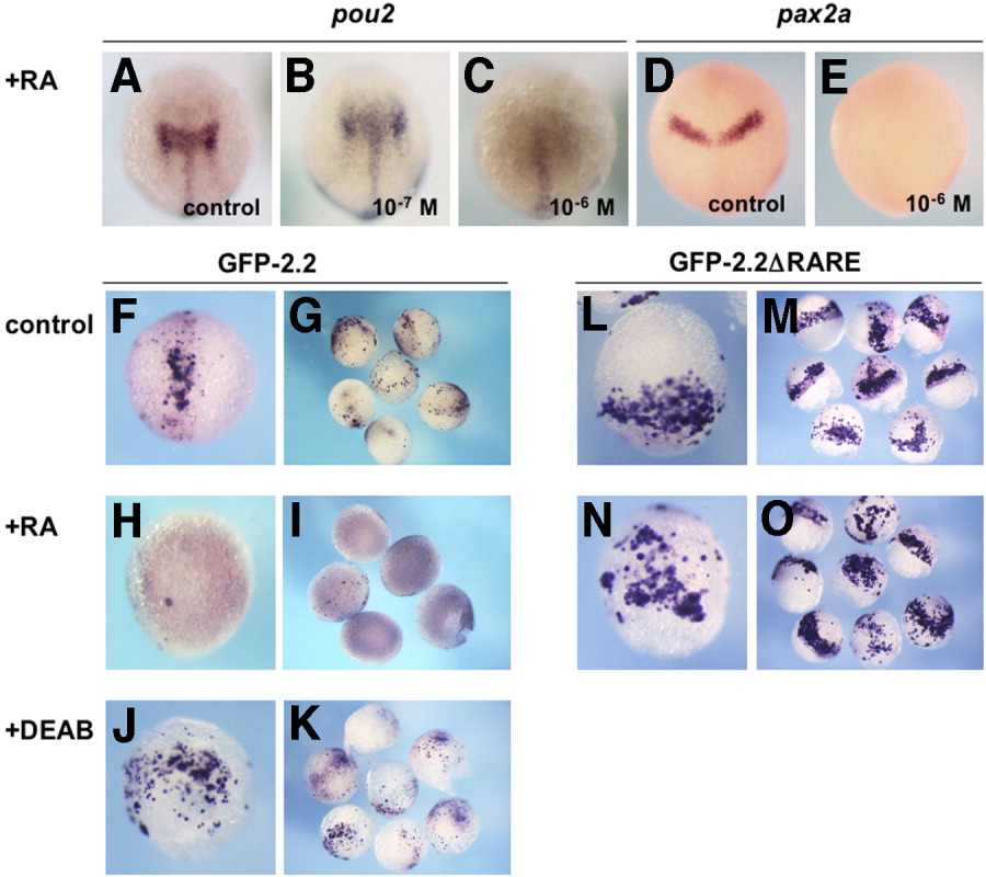
Fig. 9 Repression of pou2 and GFP-2.2 expression by retinoic acid (RA). The mRNA expression of pou2 and pax2a in uninjected embryos (A-E) and green fluorescent protein (GFP) constructs in injected embryos (F-O) was examined by whole-mount in situ hybridization. A-C: Expression of endogenous pou2 mRNA at the bud stage in the mid-hindbrain (A) was expanded by 10-7 M RA (B), whereas repressed effectively by 10-6 M RA (C). D,E: Expression of pax2a mRNA at the bud stage in the MHB region (D) was repressed by 10-6 M RA (E). F-I: Expression of GFP-2.2 in injected late gastrulae, which was seen in the mid-hindbrain region (F,G), was effectively repressed by 10-6 M RA (H,I). J,K: GFP-2.2 expression in injected embryos expanded laterally in the posterior region by diethylaminobenzaldehyde (DEAB) treatment. L,M: Expression of GFP-2.2ΔRARE in injected embryos was expanded laterally near the blastoderm margin compared with that of GFP-2.2. N,O: Expression of GFP-2.2 ΔRARE in embryos treated with 10-6 M of RA. The observed expression pattern was indistinguishable from that in untreated embryos (L,M). A-F,H,J,L,N: Dorsal views with anterior to the top. RARE, retinoic acid-responsive element.