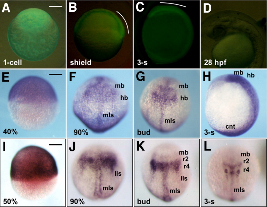
Fig. 5 Stable expression of GFP-6.5 in transgenic embryos. A-C: Expression of GFP-6.5 as visualized with green fluorescent protein (GFP) fluorescence in a fertilized egg (A), or in developing embryos at the shield (B) and three-somite (C) stages. D: GFP expression could not be detected at 28 hours postfertilization (hpf) in the head. GFP expression in the dorsal blastoderm and head regions is marked with white curves. E-H: Expression of GFP-6.5 mRNA, detected by whole-mount in situ hybridization, in embryos at 40% epiboly (E), 90% epiboly (F), bud (G), and three-somite (H) stages. I-L: Endogenous expression of pou2 at 50% epiboly (I), 90% epiboly (J), bud (K), and three-somite stages (L). A,E,I: Lateral views with animal poles to the top. B-D,H: Lateral views with anterior to the top and dorsal to the right (B,C,H) or with anterior to the left and dorsal to the top (D). F,G,J-L: Dorsal views with anterior to the top. cnt, caudal neural tube; hb, hindbrain; lls, lateral longitudinal stripe; mb, midbrain; mls, medial longitudinal stripe. Scale bar = 200 μm.
Image
Figure Caption
Figure Data
Acknowledgments
This image is the copyrighted work of the attributed author or publisher, and
ZFIN has permission only to display this image to its users.
Additional permissions should be obtained from the applicable author or publisher of the image.
Full text @ Dev. Dyn.

