
|
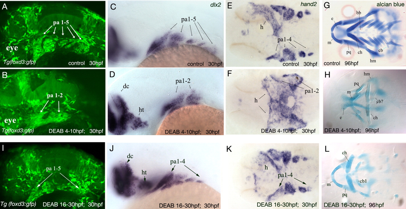
Fig. 3 DEAB treatment during gastrulation causes loss of the posterior neural crest streams and cartilages, whereas treatment post-gastrulation causes fusion of neural crest populations. A,B: Lateral views of confocal projections of GFP-positive cranial neural crest cells in Tg(foxd3:gfp) embryos. Anterior to the left. A: In 30-hpf control embryo, neural crest cells are present in pharyngeal arches 1-5. B: In embryos treated between 4-10 hpf, neural crest cells only populate the first two arches. C,D: Lateral views of 30-hpf embryos labeled with dlx2. C: dlx2 is expressed in neural crest cells in pharyngeal arches 1-5, the diencephalon, and the hypothalamus. D: Expression of dlx2 is lost in the posterior pharyngeal arches 3-5 of embryos treated during gastrulation. E: Ventral view of hand2 expression in the ventral part of pharyngeal arches 1-4 and in the developing heart tube in 30-hpf wild type embryos. F: In embryos treated during gastrulation, only the first two pharyngeal arches are labeled and heart tube morphogenesis does not occur. G,H: In embryos treated with DEAB during gastrulation, the cartilages of the mandibular and hyoid arches are present but smaller in size and misshapen. Cartilages of the posterior arches (3-7) are absent. Ventral views. I: In 30-hpf Tg(foxd3:gfp) embryos treated with DEAB between 16-30 hpf, neural crest cell have migrated into all pharyngeal arches but neural crest populations of the 4th and 5th pharyngeal arches are fused. J,K: In embryos treated post-gastrulation, dlx2 and hand2 are expressed in pharyngeal arches 1-4. L: Even though neural crest cells are present in the posterior pharyngeal arches of embryos treated with DEAB post-gastrulation, they do not differentiate into cartilage. bb, basibranchial; cb, ceratobranchial; ch, ceratohyal; dc, diencephalon; e, ethmoid plate of neurocranium; h, heart tube; hm, hyomandibula; ht, hypothalamus; m, meckel's cartilage; pa1-5, pharyngeal arches 1-5; pq, palatoquadrate.