
|
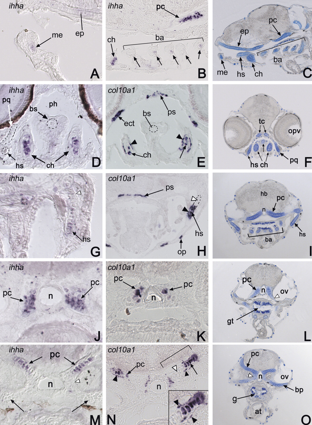
Fig. 4 ihha and col10a1 expression in the head skeleton of 6dpf larvae. In situ hybridizations using ihha (A, B, D, G, J, M) and col10a1 (E, H, K, N) probes and Alcian blue staining of cartilage cells (C, F, I, L, O) performed on 12-μm longitudinal (A-C) or transverse (D-O) cryosections of 6dpf larvae. A-C: Longitudinal sections. ihha is expressed in chondrocytes of several cartilaginous elements, such as the Meckel's (me, in A), ceratohyal (ch in B), and parachordal (pc, in B) cartilages. D-O: Cross-sections comparing ihha (left column), col10a1 (middle column) expression, and Alcian blue staining (right column) at different rostro-caudal levels (sections progress caudally). ihha and col10a1 are expressed in chondrocytes (black arrows) of the ceratohyal (ch), hyosymplectic (hs), and parachordal (pc) cartilages. Comparison with Alcian blue staining (right) positively identifies these cells as chondrocytes. In addition to cartilage cells, col10a1 is also expressed in flat cells surrounding the cartilage and several forming dermal bones (black arrowheads). White arrowheads on G, H, M, and N show chondrocytes expressing neither ihha nor col10a1, suggesting that they are less differentiated. See text for details. at, atrium; ba, branchial arches; bp, basal plate; bs, basibranchial; ch, ceratohyal; ep, ethmoid plate; ect, ectopterygoid; fb, forebrain; g, gut; hb, hindbrain; hs, hyosymplectic; me, Meckel's cartilage; n, notochord; op, opercle; opv, optic vesicle; ov, otic vesicle; pc, parachordal cartilage; ph, pharynx; ps, parasphenoid; pq, palatoquadrate; tc, trabecula.