
|
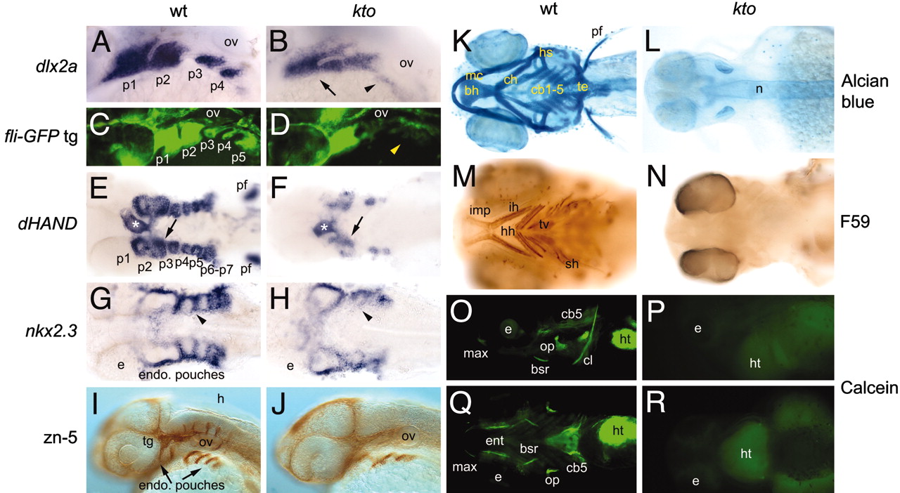
Fig. 6 Defects in the segmentation of pharyngeal arches in kto mutants. (A-D, I, J, O, and P) Lateral views, all others are ventral views. (A and B) Dlx2a expression in pharyngeal arches at 26 hpf. No distinct pharyngeal arches form (arrow), and expression in posterior arches is essentially extinguished in kto mutants (arrowhead). (C and D) TG(fli1:EGFP)y1 transgenic embryos at 31 hpf. In kto mutant embryos, p1 and p2 seem malformed and fused, whereas the more posterior arches are almost entirely lost (arrowhead). (E and F) dHAND expression in pharyngeal arches at 36 hpf. Arrows indicate separation of p1 and p2. In kto embryos, separation of p1 and p2 failed, and p3-p7 are largely absent. dHAND expression in the heart is present (white asterisk), but the pectoral fins are lost in kto.(G and H) Expression of nkx2.3 at 36 hpf visualized endodermal pouches; cells separating individual arches are lost in the mutant (arrowhead). (I and J) zn5 stains the endodermal pouches, trigeminal ganglia, and sensory neurons. (K and L) Alcian blue staining at 5 dpf. No cartilage elements are visible in the mutant. (M and N) Detection of larval muscles with F59 antibody at 4 dpf; muscles in the head are lost in the mutant. (O-R) Calcein-stained fluorescent image shows loss of bones in the head of kto embryos. b, branchyal arch; bh, basihyal; cb, ceratobranchial; ch, ceratohyal; e, eye; fb, forebrain; h, hyoid arch; hh, hyohyoideus; hs, hyosymplectic; ht, heart; ih, interhyoideus; imp, intermandibularis posterior; m, mandibular arch; mc, Meckel′s cartilage; n, notochord; ov, otic vesicle; p, pharyngeal arch; pf, pectoral fin, r, rhombomere; sh, sternohyoideus; te, teeth; tg, trigeminal ganglia; tv, transversus ventralis.