
|
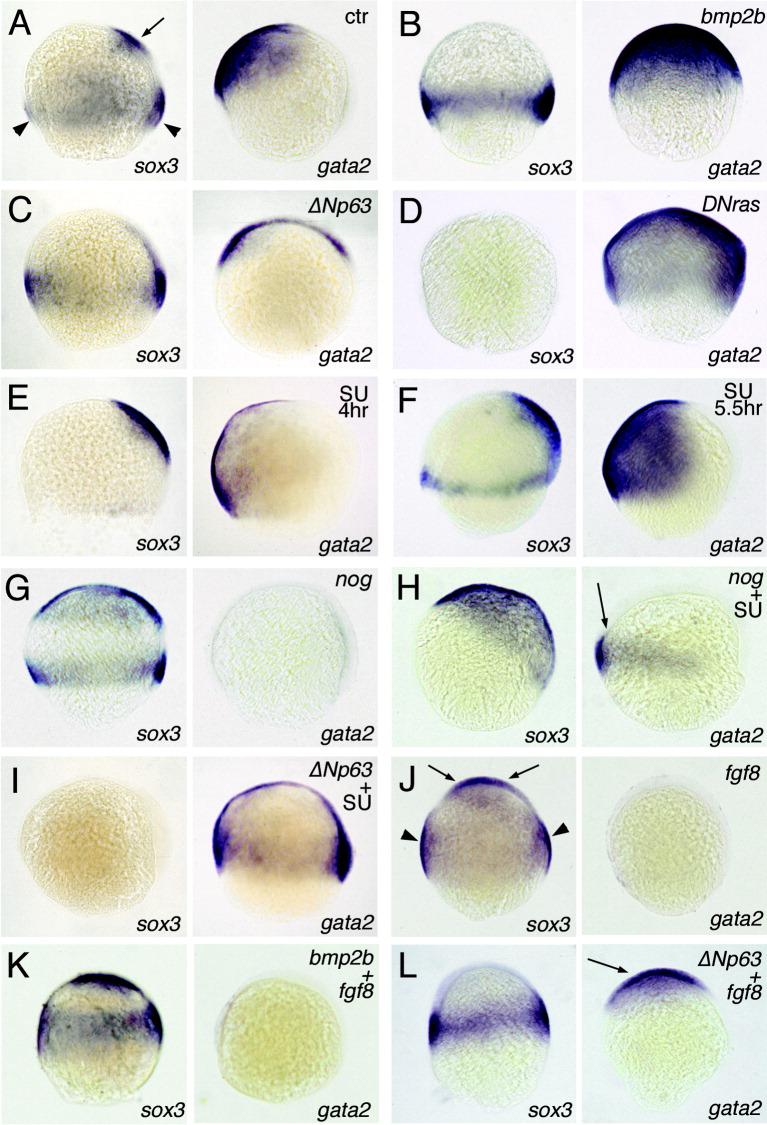
Fig. 2 Differential effects of gain or loss of fibroblast growth factor (Fgf), bone morphogenetic protein (Bmp), or ΔNp63 function on ectodermal patterning. Panels show in situ hybridizations for sox3 (neuroectoderm) on the left and for gata2 (non-neural ectoderm) on the right, 80% epiboly, dorsal to the right, animal/anterior pole up. A,J: Arrows indicate sox3 expression in anterior neuroectoderm, arrowheads sox3 expression in posterior/marginal neuroectoderm. Injected mRNAs and SU5402 treatments (SU) are indicated in the upper right corner. E,H,I: Embryos were treated with 20 μM from 4 hpf to 8 hpf, embryos in F with 20 μM SU5402 from 5.5 hpf to 8 hpf. H: Coinjection of noggin mRNA abolishes the endogenous but not the SU-induced ectopic marginal gata2 expression (indicated with an arrow). L: Coinjected ΔNp63 mRNA abolishes anterior sox3 expression, and rescues gata2 expression (indicated with an arrow), whereas it cannot block the Fgf-induced expansion of the marginal/posterior sox3 domain. Siblings of embryos shown in all panels were also stained for the anterior neural marker otx2 (Li et al., 1994) and the posterior neural marker hoxb1b (Alexandre et al., 1992), leading to consistent results (Fig. 3, and data not shown). ctr, uninjected or untreated control embryo.