
Fig. 7
- ID
- ZDB-FIG-240229-203
- Publication
- Rizvi et al., 2023 - VEGFA mRNA-LNP promotes biliary epithelial cell-to-hepatocyte conversion in acute and chronic liver diseases and reverses steatosis and fibrosis
- Other Figures
- All Figure Page
- Back to All Figure Page
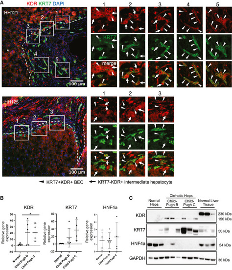
Evidence of KDR expression on BECs in human liver samples from nonalcoholic steatohepatitis (NASH) cirrhosis and alcoholic cirrhosis ESLD patients (A) Immunofluorescence images of cirrhotic liver specimens HH121 and HH125 showing fibrous septa stained for KRT7+ BECs and KDR+ sinusoidal endothelial cells. Note numerous KRT7+KDR+ BECs (arrowheads) and larger KDR+ intermediate hepatocyte-like cells (arrows) surrounding the KRT7+KDR+ BECs. (B) Relative gene expression of KDR, KRT7, and HNF4A in hepatocytes (Heps) isolated from normal (n = 5) or cirrhotic human livers scoring Child-Pugh B (n = 5) and Child-Pugh C (n = 4). (C) Western blots showing protein expression of KDR, KRT7, and HNF4A in hepatocytes isolated from normal or cirrhotic human livers Child-Pugh B (n = 3) and Child-Pugh C (n = 3) analyzed in (B). GAPDH serves as endogenous control. Numerical data are presented as mean ± SD Two-tailed Student’s t test for comparison between two groups; ∗p < 0.05. See also Figure S7. |