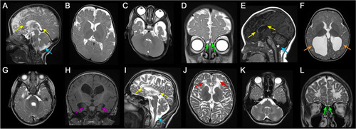
Neuroimaging findings of individuals with OGDHL pathogenic variants. Brain MRI findings of individual 1 (A-D), 6 (E–H), and 7 (I-L). Sagittal T2 (A and I) and T1 (E) weighted images showed markedly hypoplastic corpus callosum in individual 1 (A) and 7 (I) and dysplastic corpus callosum in individual 6 (E) with hypoplastic rostrum, genu, and anterior body and absent posterior body and splenium (yellow arrows). Mega cisterna magna was present in all affected individuals (A, E, and I, blue arrows), and individual 6 also had inferior vermian hypoplasia and widening of the foramen of Magendie (E, asterisk). Axial T2-weighted images (B, C, F, G, J, and K) revealed varying degrees of diffuse white matter volume loss, most severe in individual 6 (F and G) and 7 (J and K). Individual 7 also had scattered areas of leukomalacia (J, red arrows) and prominent involvement of brainstem and cerebellar white matter. Individual 6 had ventriculomegaly and colpocephaly (F, orange arrows). Coronal T2 (D and L) and T1 (H) weighted images showed hypoplastic olfactory bulbs in individual 1 (D) and 7 (L) (green arrows) and hypoplastic hippocampi in individual 6 (H, purple arrows)
|
