
FIGURE
Fig. 2
Fig. 2
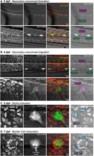
Induction of the stem cell niche in secondary neuromasts. Confocal imaging of double Tg(Eya1:eGFP)(K15:H2B-RFP) during secondary neuromast formation. (A) The forming secondary neuromast can be detected between two primary EGFP+ organs. (B) Primary neuromasts move to the ventral side while forming secondary organs migrate dorsally. (C) First epithelial cells transforming into border cells are detected in the neuromast periphery by 5dfp. (D) The neuromast has established its characteristic architecture and is surrounded by several mature border cells discernible by their elongated nuclei. |
Expression Data
Expression Detail
Antibody Labeling
Phenotype Data
Phenotype Detail
Acknowledgments
This image is the copyrighted work of the attributed author or publisher, and
ZFIN has permission only to display this image to its users.
Additional permissions should be obtained from the applicable author or publisher of the image.
Full text @ Cells Dev