
Fig. 1
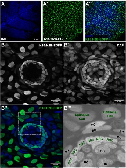
The stem cell niche in mature medaka neuromasts. (A) Low resolution confocal images of medaka juveniles - 10dpf. Neuromasts are recognised by DAPI stn (A) and by H2B-EGFP expression in Tg(K15:H2B-EGFP) (A’, A”). (B) High magnification view of a medaka mature neuromasts. The Tg(K15:H2B-EGFP) allows for visualization of epithelial cells (rounded nuclei in B, periphery; B”’) and neuromast stem cells (central, packed cells in B). Counterstain with DAPI reveals border cells (B’-B”’) with elongated nuclei wrapping the EGFP+ stem cells (NSCs) in the neuromast (B”-B”’). HC in B”’ indicates neuronal hair cells. Scale bar is 50 μm in A and 10 μm in B. |