
FIGURE
Fig. 3
- ID
- ZDB-FIG-230906-15
- Publication
- Arias-Alpizar et al., 2022 - Phase-separated Liposomes Hijack Endogenous Lipoprotein Transport and Metabolism Pathways to Target Subsets of Endothelial Cells in vivo
- Other Figures
- All Figure Page
- Back to All Figure Page
Fig. 3
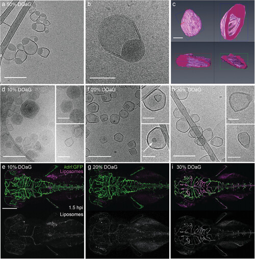
Cryo-TEM of PAP3 liposomes characterized by phase separation correlates with the bECs targeting in zebrafish larvae (≈78 hpf). a,b) CryoTEM of PAP3 (50–50 mol% DOaG-DSPC) liposomes and c) 3D model of a representative PAP3 liposome reconstructed based on the electron density derived from cryo-electron tomography, demonstrating the whole body and different plane sections of the particle. CryoTEM and biodistribution (in a Tg(kdrl:GFP) zebrafish embryo at 1.5 hpi) of liposomes containing DSPC and d,e) DOaG 10 mol%, f,g) 20 mol%, and h,i) 30 mol%. Liposomes (5 mM, 0.2% mol DOPE-LR) described in all panels formulated by ethanol injection except panels (b) and (c) – formulated by extrusion. Scale bars: 200 nm and 100 nm for higher magnification insets for CryoTEM, 100 nm for 3D reconstruction, and 200 µm for dorsal zebrafish view.
|
Expression Data
Expression Detail
Antibody Labeling
Phenotype Data
Phenotype Detail
Acknowledgments
This image is the copyrighted work of the attributed author or publisher, and
ZFIN has permission only to display this image to its users.
Additional permissions should be obtained from the applicable author or publisher of the image.
Full text @ Adv. Healthc. Mater.