FIGURE

FIGURE 1

ID
ZDB-FIG-230625-4
Publication
Liu et al., 2023 - Retinal degeneration in rpgra mutant zebrafish
Other Figures
All Figure Page
Back to All Figure Page
FIGURE 1

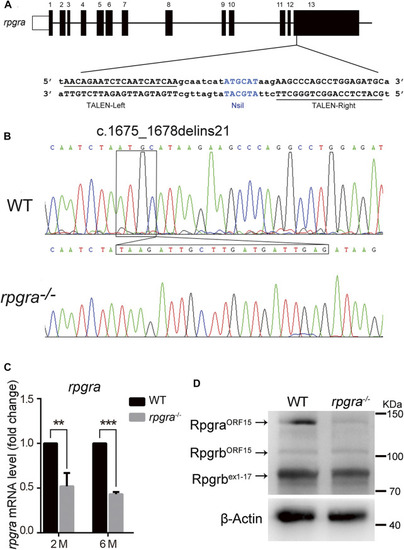
Generation of the rpgra mutant zebrafish. (A) The 13 exons of zebrafish rpgra are shown with the left and right arms of the TALEN binding sequences underlined. TALEN-L and TALEN-R, the left and right arms of the TALENs binding sequences; the NsiI restriction site in the spacer region is used for mutation detection. (B) Sequencing of the c.1675_1678delinsTAAGATTGCTTGATGATTGAG rpgra mutation in homozygous zebrafish. The DNA base change was indicated with a box. (C)rpgra mRNA levels in 2- and 6-month-old WT and rpgra−/− zebrafish eyes detected by quantitative PCR. Actb1 served as endogenous control. The result was shown as mean ± SD. **, 0.001 < p < 0.01; ***, p < 0.001. (D) Rpgra protein levels of WT and rpgra−/− zebrafish at 2mpf were revealed by Western blot using the anti-RPGR antibody.

Expression Data

Expression Detail
Antibody Labeling
Phenotype Data

Phenotype Detail
Acknowledgments
This image is the copyrighted work of the attributed author or publisher, and ZFIN has permission only to display this image to its users. Additional permissions should be obtained from the applicable author or publisher of the image. Full text @ Front Cell Dev Biol