
Fig. 3
- ID
- ZDB-FIG-230403-12
- Publication
- Lin et al., 2022 - Optokinetic set-point adaptation functions as an internal dynamic calibration mechanism for oculomotor disequilibrium
- Other Figures
- All Figure Page
- Back to All Figure Page
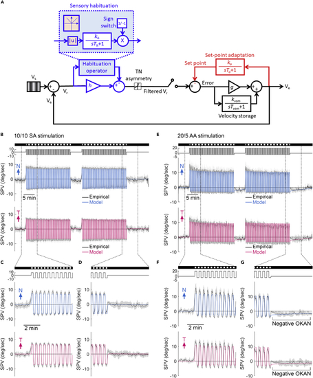
Optokinetic set-point adaptation model predicts the recorded SPV in larval zebrafish (A) The mathematical model of set-point adaptation. Retinal slip velocity (Vr) is the difference between the stimulus velocity (Vs) and the eye velocity (Ve). During habituation, Vr is rectified and filtered with a time constant of Th and gain of kh (blue shaded area), and then scaled with h, to give a filtered Vr. In the sensory habituation operator, |u|, which produces the absolute value of the input (orange shaded area), together with a sign switch of output lead to a continuous habituating effect regardless of stimulus direction. The filtered Vr passes through a nonlinear gain (T-N gain) that captures the T-N asymmetry. The error between the filtered Vr and the set point is scaled by oculomotor gain (g) and added to the velocity storage mechanism (VSM) to control Ve. VSM contains a leaky integrator with a time constant of Tvsm and a gain of kvsm that contributes to the eye velocity. Ve is then integrated with a time constant of Ta and a gain of ka to adjust the set point. (B–G) The model predicted population-median SPV (colored lines) superimposed on the median ± median absolute deviation (black line with gray shadow) of empirical SPVs during 10/10 symmetric alternating stimulations (SA, n = 29) (B–D) and 20/5 asymmetric alternating stimulations (AA, n = 18) (E–G). Considering the starting direction of alternating stimulation was randomized, we aligned the first stimulus direction as positive instead of left or right. Therefore, the plots show one eye started with a nasalward movement (blue trace) and another eye started with a temporalward movement (red trace). The stimulus image pattern (darkness or gratings) and the stimulus velocity are shown on the top as horizontal bars and lines, respectively. |