Figure 8
- ID
- ZDB-FIG-230402-8
- Publication
- Streiff et al., 2023 - Susceptibility and Permissivity of Zebrafish (Danio rerio) Larvae to Cypriniviruses
- Other Figures
- All Figure Page
- Back to All Figure Page
|
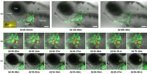
Frames from timelapse video of CyHV-3 EGFP infection in zebrafish larvae from 2–3 dpi (Video S1). The video represents overlay of brightfield/transmission and EGFP fluorescence (green). Time postinfection (in days, hours, and minutes) is indicated under each frame. (a) Entire field of view from light-sheet microscopy. For the purposes of visual orientation, identifiable anatomical features and corresponding locations within larvae body (inset image) are indicated in the first panel. Images show that the infection is primarily localized around the inoculation site (red square), and a decrease in viral levels from 2.5–3 dpi. Scale bars = 100 µm. (b) Enlarged images of the area within red square in (a), representing key examples of apoptosis-like death occurring among large numbers of infected cells (red circles) around the inoculation site, with such events primarily characterized by blebbing followed by the appearance of cell debris resembling apoptotic bodies (c) Key example of highly motile infected cell (highlighted with yellow circle), migrating away from the site of inoculation. |

