
Fig. 1
- ID
- ZDB-FIG-230225-16
- Publication
- Fang et al., 2021 - A membrane arm of mitochondrial complex I sufficient to promote respirasome formation
- Other Figures
- All Figure Page
- Back to All Figure Page
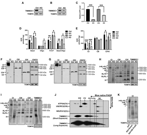
Figure 1. Loss of TIMMDC1 leads to the accumulation of respirasome subcomplex (A and B) Western blot (WB) analysis of the steady-state levels of TIMMDC1 in HEK293T cells with KO of TIMMDC1 (A), KD of TIMMDC1 (B), and paired control cells (Ctrl). TOM70 was used as an internal control. (C) Cellular ATP content in HEK293T cells with KO and KD of TIMMDC1 and paired control cells (Ctrl). (n = 3) (D) Cell respiration was determined by measuring the oxygen consumption rate (OCR) in HEK293T cells with KO and KD of TIMMDC1 and in paired control cells (Ctrl). Endogenous respiration is presented as follows: basal, basal-cell respiration; oligo, the non-phosphorylating proton leaked respiration measured in the presence of oligomycin (2 μg/mL); FCCP, un-coupling respiration measured in the presence of carbonyl cyanide 4-(trifluoromethoxy)phenylhydrazone (2.5 μM); basal-oligo, absolute respiration of ADP to ATP phosphorylation. (n = 3) (E) Respiratory complex-dependent OCR in HEK293T cells with KO and KD of TIMMDC1 and in paired control cells (Ctrl). CI, respiration in the presence of malate and glutamate. CII, respiration in the presence of succinate. CI+CII, respiration in the presence of malate, glutamate, and succinate. (n = 3) (F and G) Steady-state levels of OXPHOS complexes were analyzed in HEK293T cells with KO (F) and KD (G) of TIMMDC1 and in paired control cells (Ctrl). N-dodecyl β-d-maltoside (DDM) solubilized protein was separated by 3.5%–16% blue-native PAGE. OXPHOS complexes were detected by immunoblotting with antibodies against proteins as indicated (CI, NDUFA13; CII, SDHA; CIII, UQCRC2; CIV, MT-CO1; CV, ATP5A). Complexes I–V are abbreviated as CI–V. TOM70 was used as an internal control. (H and I) Respiratory supercomplexes were analyzed in HEK293T cells with KO (H) and KD (I) of TIMMDC1 and in paired control cells (Ctrl). Digitonin-solubilized protein was separated by 3%–11% blue-native PAGE. Respiratory supercomplexes were immunoblotted with antibodies as indicated (CI, NDUFB6; CIII, UQCRC2; CIV, MT-CO1). TOM70 was used as an internal control. (J) 2D blue-native PAGE/tricine SDS-PAGE and immunoblotting analysis of HEK293T cells. Digitonin-solubilized protein was separated by 1D 3%–11% blue-native PAGE, followed by 2D tricine SDS-PAGE. OXPHOS complexes were immunoblotted with anti-CI (NDUFB6), -CIII (UQCRC2), -CV (ATP5A), and -TIMMDC1 antibodies. (K) Blue-native PAGE and immunoblotting analysis of HEK293T cells (Ctrl), TIMMDC1−/− cells with empty vector (KO+vector), and TIMMDC1−/− cells with re-expression of FLAG-tagged TIMMDC1 (KO+OE-FLAG; OE [overexpression]). Digitonin-solubilized protein was separated by 3%–11% blue-native PAGE and then immunoblotted with the anti-NDUFB6 (CI) antibody. TOM70 was used as an internal control. Quantitative data were generated from three independent experiments. Data are presented as the means ± SEM. ∗p < 0.05, ∗∗p < 0.01, ∗∗∗p < 0.001. |