|
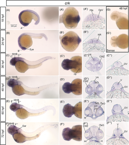
crk mRNA is expressed in the developing nervous system in zebrafish. A–F, Lateral whole mount and (A′–F′) dorsal whole mount in situ hybridization embryos for crk expression at stages 18 hpf–72 hpf. Twenty micron transverse sections through the forebrain (A″–F″) and hindbrain (C″–F″) of in situ embryos at the lines indicated in (A–F). G, Lateral and (G′) dorsal images of sense control probe on 48 hpf embryos. Scale bar at 250 μm (A) and 75 μm (A″). CMZ, ciliary marginal zone; DT, dorsal thalamus; FB, fin bud; FBr, forebrain; H, heart; lTeO, lateral optic tectum; MHB, midbrain-hindbrain boundary; NR, neural retina; oc, optic chiasm; OpV, optic vesicle; OV, otic vesicle; PT, posterior tuberculum; R, retina; TeO, optic tectum; V, ventricle; V4, fourth ventricle
|