
Figure 2
- ID
- ZDB-FIG-230124-184
- Publication
- Porcino et al., 2023 - Potential Neuroprotective Role of Calretinin-N18 and Calbindin-D28k in the Retina of Adult Zebrafish Exposed to Different Wavelength Lights
- Other Figures
- All Figure Page
- Back to All Figure Page
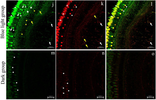
Calbindin-D28K/Calretinin-N18 immunostaining in zebrafish (Danio rerio) retina, approximately 4-months-old. (a–c) Calbindin-D28K/Calretinin-N18 immunostaining in control group (natural photoperiod conditions) (a) CaB-D28K-immunoreactivity in the cytoplasmic prolongations of the cells of the retinal pigment epithelium (RPE) (chevron arrows), in the cones and rods of the photoreceptors layer (bold arrows). (b) CaR-N18 immunoreactivity in the outer plexiform layer (OPL) (asterisks), in the soma of a subpopulation of amacrine cells (arrowheads) and bipolar cells (arrows), in the inner plexiform layer (IPL) (stars), and in the soma of ganglion cells (GCs) (yellow arrows). (c) Merge: absence of Calbindin-D28K/Calretinin-N18 colocalization. (d–f) Calbindin-D28K/Calretinin-N18 immunostaining in white light group (emission 34.8% of 400–500 nm). (d) CaB-D28K-immunoreactivity in the cytoplasmic prolongations of the cells of the retinal pigment epithelium (RPE) (chevron arrows), in the outer segments of cones (bold arrows), and inner segment of cones (yellow arrowheads) and in the rods (yellow bold arrows), in the outer plexiform layer (OPL) (arrows), in the bipolar cells (yellow arrows), in the inner plexiform layer (IPL) (stars), and in the ganglion cells and their axons (GC) (asterisks). (e) Calretinin-N18 immunoreactivity in the inner segments of cones (bold arrows), in the rods (arrows), in the innermost layer of the OPL (yellow arrows), in the innermost layer of the IPL (stars) and in the GCs and their axons (asterisks). (f) Merge: Calbindin-D28K/Calretinin-N18 colocalization in inner segments of cones (bold arrows), in the rods (arrows), in the innermost layer of IPL (stars) and OPL (yellow arrows) and in the GCs and their axons (asterisks). (g–i) Calbindin-D28K/Calretinin-N18 immunostaining in white–blue light group (emission 54.6% of 400–500 nm). (g) Calbindin-D28K immunoreactivity in retinal pigment epithelium (RPE) into the cytoplasmic prolongations of its cells (chevron arrow), in the inner (bold arrows) and outer segment of cones (arrows heads), in the rods (yellow bold arrows), in the innermost layer of OPL (arrows), in the bipolar cells (yellow arrows) and in the IPL (stars). (h) Calretinin-N18 immunoreactivity in the inner segments of cones (bold arrows), in the rods (yellow bold arrows), in the innermost layer of OPL (arrows), in the BCs (yellow arrows), in a subpopulation of amacrine cells (arrowheads), in the innermost layer of OPL (stars), and in the GCs (asterisk). (i) Merge: Calbindin-D28K/Calretinin-N18 colocalization in the photoreceptor layer (bold arrows), in the innermost layer of OPL (arrows), in the BCs (yellow arrows) and in the innermost layer of IPL (stars). (j–l) Calbindin-D28K/Calretinin-N18 immunostaining in blue light group (emission 84.3% of 400–500 nm). (m) Calbindin-D28K immunoreactivity in the outer (arrowheads) and inner (bold arrows) segment of cones and in the rods (asterisks), in the OPL (stars), in some amacrine cells (yellow bold arrows), in the BCs (yellow arrows), and GCs (arrows). (n) Calretinin-N18 immunoreactivity in the inner segments of cones (bold arrows) and rods (asterisks), in a subpopulation of amacrine cells (yellow arrows), GCs, and their axons (arrows). (o) Merge: Calbindin-D28K/Calretinin-N18 colocalization in the inner segments of cones (bold arrows) and in the rods (asterisks) and in some GCs and their axons (arrows). (m–o) Sample kept in dark conditions. (m) Low CaB-D28K immunoreactivity and (n) low CaR-N18 immunoreactivity in the photoreceptors layer. No immunostaining to Calbindin-D28K (a) and Calretinin-N18 (b) in the other retinal layers. (o) Merge: no immunostaining to Calbindin-D28K/Calretinin-N18. Magnification 40× (a–o). |