
- Title
-
Potential Neuroprotective Role of Calretinin-N18 and Calbindin-D28k in the Retina of Adult Zebrafish Exposed to Different Wavelength Lights
- Authors
- Porcino, C., Briglia, M., Aragona, M., Mhalhel, K., Laurà, R., Levanti, M., Abbate, F., Montalbano, G., Germanà, G., Lauriano, E.R., Meduri, A., Vega, J.A., Germanà, A., Guerrera, M.C.
- Source
- Full text @ Int. J. Mol. Sci.
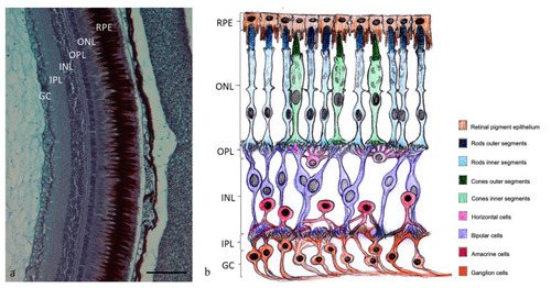
(a) Retina of adult zebrafish: RPE, retinal pigment epithelium; ONL, outer nuclear layer; OPL, outer plexiform layer; INL, inner nuclear layer; IPL, inner plexiform layer; GC, ganglion cell layer; Alcian Blue-PAS staining. Magnification 20×. (b) Graphical representation of the cell layers into the retina. |
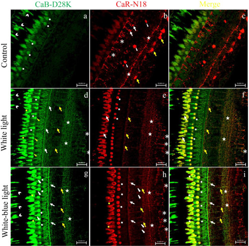
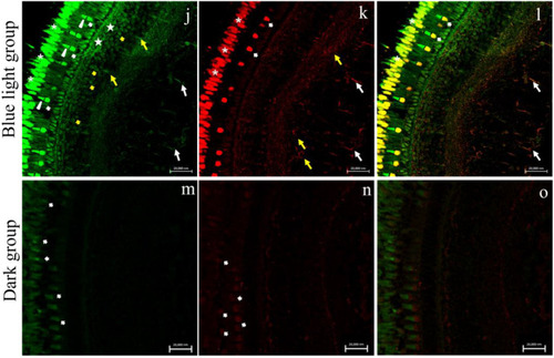
Calbindin-D28K/Calretinin-N18 immunostaining in zebrafish (Danio rerio) retina, approximately 4-months-old. (a–c) Calbindin-D28K/Calretinin-N18 immunostaining in control group (natural photoperiod conditions) (a) CaB-D28K-immunoreactivity in the cytoplasmic prolongations of the cells of the retinal pigment epithelium (RPE) (chevron arrows), in the cones and rods of the photoreceptors layer (bold arrows). (b) CaR-N18 immunoreactivity in the outer plexiform layer (OPL) (asterisks), in the soma of a subpopulation of amacrine cells (arrowheads) and bipolar cells (arrows), in the inner plexiform layer (IPL) (stars), and in the soma of ganglion cells (GCs) (yellow arrows). (c) Merge: absence of Calbindin-D28K/Calretinin-N18 colocalization. (d–f) Calbindin-D28K/Calretinin-N18 immunostaining in white light group (emission 34.8% of 400–500 nm). (d) CaB-D28K-immunoreactivity in the cytoplasmic prolongations of the cells of the retinal pigment epithelium (RPE) (chevron arrows), in the outer segments of cones (bold arrows), and inner segment of cones (yellow arrowheads) and in the rods (yellow bold arrows), in the outer plexiform layer (OPL) (arrows), in the bipolar cells (yellow arrows), in the inner plexiform layer (IPL) (stars), and in the ganglion cells and their axons (GC) (asterisks). (e) Calretinin-N18 immunoreactivity in the inner segments of cones (bold arrows), in the rods (arrows), in the innermost layer of the OPL (yellow arrows), in the innermost layer of the IPL (stars) and in the GCs and their axons (asterisks). (f) Merge: Calbindin-D28K/Calretinin-N18 colocalization in inner segments of cones (bold arrows), in the rods (arrows), in the innermost layer of IPL (stars) and OPL (yellow arrows) and in the GCs and their axons (asterisks). (g–i) Calbindin-D28K/Calretinin-N18 immunostaining in white–blue light group (emission 54.6% of 400–500 nm). (g) Calbindin-D28K immunoreactivity in retinal pigment epithelium (RPE) into the cytoplasmic prolongations of its cells (chevron arrow), in the inner (bold arrows) and outer segment of cones (arrows heads), in the rods (yellow bold arrows), in the innermost layer of OPL (arrows), in the bipolar cells (yellow arrows) and in the IPL (stars). (h) Calretinin-N18 immunoreactivity in the inner segments of cones (bold arrows), in the rods (yellow bold arrows), in the innermost layer of OPL (arrows), in the BCs (yellow arrows), in a subpopulation of amacrine cells (arrowheads), in the innermost layer of OPL (stars), and in the GCs (asterisk). (i) Merge: Calbindin-D28K/Calretinin-N18 colocalization in the photoreceptor layer (bold arrows), in the innermost layer of OPL (arrows), in the BCs (yellow arrows) and in the innermost layer of IPL (stars). (j–l) Calbindin-D28K/Calretinin-N18 immunostaining in blue light group (emission 84.3% of 400–500 nm). (m) Calbindin-D28K immunoreactivity in the outer (arrowheads) and inner (bold arrows) segment of cones and in the rods (asterisks), in the OPL (stars), in some amacrine cells (yellow bold arrows), in the BCs (yellow arrows), and GCs (arrows). (n) Calretinin-N18 immunoreactivity in the inner segments of cones (bold arrows) and rods (asterisks), in a subpopulation of amacrine cells (yellow arrows), GCs, and their axons (arrows). (o) Merge: Calbindin-D28K/Calretinin-N18 colocalization in the inner segments of cones (bold arrows) and in the rods (asterisks) and in some GCs and their axons (arrows). (m–o) Sample kept in dark conditions. (m) Low CaB-D28K immunoreactivity and (n) low CaR-N18 immunoreactivity in the photoreceptors layer. No immunostaining to Calbindin-D28K (a) and Calretinin-N18 (b) in the other retinal layers. (o) Merge: no immunostaining to Calbindin-D28K/Calretinin-N18. Magnification 40× (a–o). |
Calbindin-D28K/Calretinin-N18 immunostaining in zebrafish (Danio rerio) retina, approximately 4-months-old. (a–c) Calbindin-D28K/Calretinin-N18 immunostaining in control group (natural photoperiod conditions) (a) CaB-D28K-immunoreactivity in the cytoplasmic prolongations of the cells of the retinal pigment epithelium (RPE) (chevron arrows), in the cones and rods of the photoreceptors layer (bold arrows). (b) CaR-N18 immunoreactivity in the outer plexiform layer (OPL) (asterisks), in the soma of a subpopulation of amacrine cells (arrowheads) and bipolar cells (arrows), in the inner plexiform layer (IPL) (stars), and in the soma of ganglion cells (GCs) (yellow arrows). (c) Merge: absence of Calbindin-D28K/Calretinin-N18 colocalization. (d–f) Calbindin-D28K/Calretinin-N18 immunostaining in white light group (emission 34.8% of 400–500 nm). (d) CaB-D28K-immunoreactivity in the cytoplasmic prolongations of the cells of the retinal pigment epithelium (RPE) (chevron arrows), in the outer segments of cones (bold arrows), and inner segment of cones (yellow arrowheads) and in the rods (yellow bold arrows), in the outer plexiform layer (OPL) (arrows), in the bipolar cells (yellow arrows), in the inner plexiform layer (IPL) (stars), and in the ganglion cells and their axons (GC) (asterisks). (e) Calretinin-N18 immunoreactivity in the inner segments of cones (bold arrows), in the rods (arrows), in the innermost layer of the OPL (yellow arrows), in the innermost layer of the IPL (stars) and in the GCs and their axons (asterisks). (f) Merge: Calbindin-D28K/Calretinin-N18 colocalization in inner segments of cones (bold arrows), in the rods (arrows), in the innermost layer of IPL (stars) and OPL (yellow arrows) and in the GCs and their axons (asterisks). (g–i) Calbindin-D28K/Calretinin-N18 immunostaining in white–blue light group (emission 54.6% of 400–500 nm). (g) Calbindin-D28K immunoreactivity in retinal pigment epithelium (RPE) into the cytoplasmic prolongations of its cells (chevron arrow), in the inner (bold arrows) and outer segment of cones (arrows heads), in the rods (yellow bold arrows), in the innermost layer of OPL (arrows), in the bipolar cells (yellow arrows) and in the IPL (stars). (h) Calretinin-N18 immunoreactivity in the inner segments of cones (bold arrows), in the rods (yellow bold arrows), in the innermost layer of OPL (arrows), in the BCs (yellow arrows), in a subpopulation of amacrine cells (arrowheads), in the innermost layer of OPL (stars), and in the GCs (asterisk). (i) Merge: Calbindin-D28K/Calretinin-N18 colocalization in the photoreceptor layer (bold arrows), in the innermost layer of OPL (arrows), in the BCs (yellow arrows) and in the innermost layer of IPL (stars). (j–l) Calbindin-D28K/Calretinin-N18 immunostaining in blue light group (emission 84.3% of 400–500 nm). (m) Calbindin-D28K immunoreactivity in the outer (arrowheads) and inner (bold arrows) segment of cones and in the rods (asterisks), in the OPL (stars), in some amacrine cells (yellow bold arrows), in the BCs (yellow arrows), and GCs (arrows). (n) Calretinin-N18 immunoreactivity in the inner segments of cones (bold arrows) and rods (asterisks), in a subpopulation of amacrine cells (yellow arrows), GCs, and their axons (arrows). (o) Merge: Calbindin-D28K/Calretinin-N18 colocalization in the inner segments of cones (bold arrows) and in the rods (asterisks) and in some GCs and their axons (arrows). (m–o) Sample kept in dark conditions. (m) Low CaB-D28K immunoreactivity and (n) low CaR-N18 immunoreactivity in the photoreceptors layer. No immunostaining to Calbindin-D28K (a) and Calretinin-N18 (b) in the other retinal layers. (o) Merge: no immunostaining to Calbindin-D28K/Calretinin-N18. Magnification 40× (a–o). |
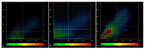
Scatter plot of CaB-D28K/CaR-N18 colocalization in different experimental groups: (a) white-light experimental group; (b) white/blue-light experimental group; (c) blue-light experimental group. Zen 2011 (LSM 700 Zeiss software, Germany). |
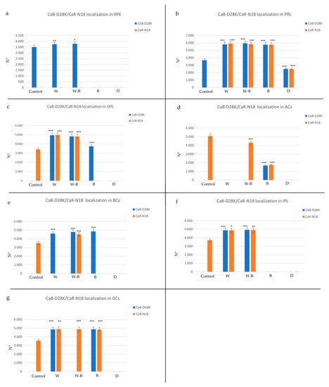
Graphical representation of immunopositivity of: (a) retinal pigment epithelium (RPE), (b) photoreceptor layer (PRL), (c) outer plexiform layer (OPL), (d) amacrine cells (ACs), (e) inner plexiform layer (IPL), (f) bipolar cells (BCs), (g) ganglion cells (GCs) detected by Calbindin (CaB-D28K) and Calretinin (CaR-18N) in different experimental groups. Experimental conditions: control (natural photoperiod), white light with a 34.8% of blue light emission (W), white–blue light with a 54.6% of blue light emission (WB), blue light with an 84.3% of blue light emission (B) and darkness (D). The statistical analysis shows a different distribution pattern of the two proteins between the experimental groups compared to the control. Data represent the average of measurements from the ten sections from each treatment. Statistical significance: *** p < 0.001, ** p < 0.01, * p < 0.05. |
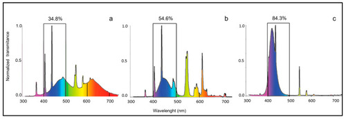
Spectral distribution of the illumination sources. (a) white light, (b) white–blue light, (c) blue light. The percentage of blue light emission for each experiment is indicated. |