
Figure 3
- ID
- ZDB-FIG-230123-18
- Publication
- Sabharwal et al., 2022 - Genetic therapy in a mitochondrial disease model suggests a critical role for liver dysfunction in mortality
- Other Figures
-
- Figure 1
- Figure 2
- Figure 2 - figure supplement 1
- Figure 2 - figure supplement 2
- Figure 2 - figure supplement 3
- Figure 3
- Figure 3 - figure supplement 1
- Figure 4
- Figure 4 - figure supplement 1
- Figure 5
- Figure 5 - figure supplement 1
- Figure 5 - figure supplement 2
- Figure 6
- Figure 7
- Figure 7 - figure supplement 1
- Figure 8
- All Figure Page
- Back to All Figure Page
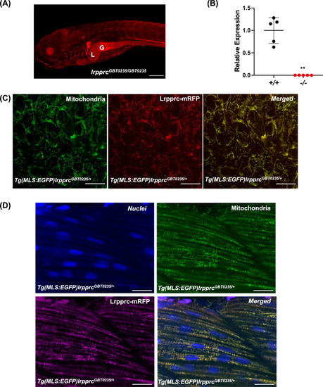
Spatiotemporal expression of Lrpprc-mRFP in GBT0235 mutants.(A) Representative images of 6 dpf lrpprcGBT0235/GBT0235 with RFP expression in the liver and gut (magnification-5×; scale-bar: 200 µm). (B) Relative expression of lrpprc transcript in lrpprc+/+ and homozygous mutant larvae, lrpprc-/- (P-value = 0.0015). p-value was determined using the unpaired t-test. Each data point represents a biological replicate (N=5) (Figure 3—source data 1). (C) Mitochondrial network marked by EGFP in the caudal fin was observed to be overlapping with truncated Lrpprc:mRFP fusion protein in 2 dpf Tg(MLS:EGFP)lrpprcGBT0235/+ embryo (scale bar: 15 µm). (D) RFP from truncated Lrpprc:mRFP fusion protein was observed to be overlapped with EGFP in mitochondria present in myocytes of skeletal muscle region in 2 dpf Tg(MLS:EGFP)lrpprcGBT0235/+ embryo injected with NLS:TagBFP RNA. Nuclei were marked by TagBFP protein (scale-bar: 15 µm). |
| Genes: | |
|---|---|
| Fish: | |
| Anatomical Terms: | |
| Stage Range: | Long-pec to Day 6 |