
Fig. 1
- ID
- ZDB-FIG-230120-28
- Publication
- Munjal et al., 2021 - Extracellular hyaluronate pressure shaped by cellular tethers drives tissue morphogenesis
- Other Figures
- All Figure Page
- Back to All Figure Page
In toto imaging of the developing zebrafish inner ear reveals multi-scale dynamics during semicircular canal morphogenesis
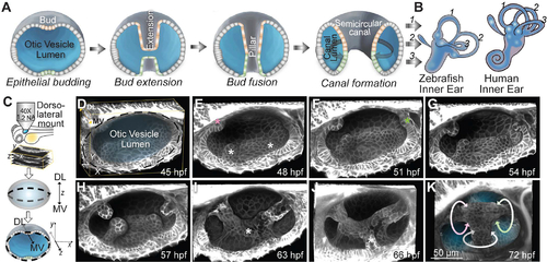
(A) Illustrations showing the formation of a SCC. (B) Illustrations of the inner ears from adult zebrafish and human comparing the conserved structures of the SCCs. Anterior, posterior and lateral SCCs are labelled as 1, 2 and 3 respectively. (C) Workflow for zebrafish embryo mounting, image acquisition and visualization. (D-K) 3D rendered otic vesicles (OVs) at select time points using Tg(βActin:membrane-Citrine). Anterior to the left and dorso-lateral (DL) into plane of view. Lateral, anterior and posterior buds are marked by white, pink and green asterisks respectively. The buds extend (G and H), and fuse (I). Ventro-lateral and ventral buds form and extend (marked by white asterisks) (I and J). Bud fusion demarcates the hubs for the anterior, posterior and lateral SCC (marked by pink, green and white circular arrows respectively) (K). Scale bar, 50 μm. |
Reprinted from Cell, 184, Munjal, A., Hannezo, E., Tsai, T.Y., Mitchison, T.J., Megason, S.G., Extracellular hyaluronate pressure shaped by cellular tethers drives tissue morphogenesis, 6313-6325.e18, Copyright (2021) with permission from Elsevier. Full text @ Cell