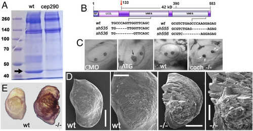
Otolithic Cochlin deficiency in mutants of cilia genes and cochlin mutant phenotype. (A) Otolithic proteins from wild-type and cep290sa1383 homozygotes separated on a polyacrylamide gel and visualized with Coomassie blue staining. The arrow indicates Cochlin band. (B) Domain structure of the cochlin gene. LCCL, Limulus factor C, Cochlin, Lgl1 domain; vWF, von Willebrand Factor domains. Deletions in CRISPR-induced mutants are indicated. The antiserum against Cochlin was made against the C terminus (amino acids 390 to 553) (C) Otolith morphology in morphant and cochsh536 mutant embryos as indicated. ATG-targeted morpholino (ATG) was used. Shown are side views of otic vesicles in live embryos, the arrows point at otoliths that are abnormal. (D) Scanning electron microscopy images of typical otoliths from wild-type and coch sh536 mutant adult at 7 mo of age (Scale bars, overview 300 μm, blow up 150 μm) (n = 3). (E) Example of cobalt nitrate staining of utricular otoliths from wild-type and coch sh536 mutant at 7 mo (n = 3).