
Fig. 7
- ID
- ZDB-FIG-230112-7
- Publication
- Anderson et al., 2021 - Tbx5a and Tbx5b paralogues act in combination to control separate vectors of migration in the fin field of zebrafish
- Other Figures
- All Figure Page
- Back to All Figure Page
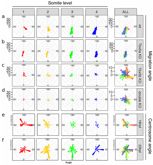
Cellular movements are randomized in Tbx5b and double knock-down embryos.
The rose diagrams in a-d represent the angle of migration with respect to the ML axis. Angles are divided along the AP axis, with cells adjacent to somite 1 in red, cells adjacent to somite 2 in yellow, cells adjacent to somite 3 in green and cells adjacent to somite 4 in blue. (a) Wildtype embryos show a directed migration, with different angles of migration for somites 1-4. (b) Tbx5a knock-down embryos display equivalent angles of migration across the AP axis. (c) Tbx5b knock-down embryos show wt-like angles of migration along the AP axis, but randomized ML movements. (d) Double knock-down embryos display both AP and ML changes to the angles of cell migration across all positions. (e-f) Angle of xCentrin::RFP expression with respect to the nucleus in the cells of the wt fin field. (e) In 18 hpf embryos, the angle of xCentrin::RFP expression with respect to the nucleus displayed no significant bias in angle. (f) At 22 hpf, the angle of xCentrin::RFP with respect to the nucleus displayed no significant bias in angle. |
Reprinted from Developmental Biology, 481, Boyle-Anderson, E.A.T, Mao, Q., Ho, R.K., Tbx5a and Tbx5b paralogues act in combination to control separate vectors of migration in the fin field of zebrafish, 201-214, Copyright (2021) with permission from Elsevier. Full text @ Dev. Biol.