
FIG 1
- ID
- ZDB-FIG-221226-139
- Publication
- Cuvry et al., 2022 - The Role of Histo-Blood Group Antigens and Microbiota in Human Norovirus Replication in Zebrafish Larvae
- Other Figures
- All Figure Page
- Back to All Figure Page
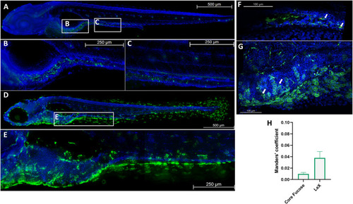
Lewis X (Lex) and core fucose are expressed in 5-days post fertilization (dpf) zebrafish larvae. (A to C) Whole-mount immunohistochemistry staining in 5 dpf larvae of core fucose stained with fluorescein isothiocyanate (FITC)-labeled A. aurantia lectin (AAL, green) and counterstained with Hoechst 33342 (blue) at ×10 (A) and ×20 (B) magnification in the intestinal bulb and ×20 magnification along the posterior intestine (C). (D, E) Whole-mount immunohistochemistry staining in 5 dpf larvae of LeX stained with anti-LeX primary antibody (green) and Hoechst 33342 (blue) at ×10 (D) and ×20 (E) magnification, representative image of LeX staining along the intestinal tract. (F, G) Whole-mount immunohistochemistry staining of human norovirus (HuNoV)-infected zebrafish larvae at ×20 magnification costained with an anti-VP1 antibody (red) and FITC-AAL (F) or LeX (G), with Hoechst 33342 as counterstaining. White arrows, colocalizing viral particles. (H) Tresholded Manders’ colocalization coefficient of HuNoV VP1 with fucose residues in the gastrointestinal tract of 5 dpf larvae (n = 5). Tresholded Manders’ coefficients are calculated with Imaris colocalization software. For panels A to E, adjacent tile pictures were merged together using the mosaic merge function of the LAS X software. |