
FIGURE
Figure 4
- ID
- ZDB-FIG-221211-181
- Publication
- Drummond et al., 2022 - osr1 Maintains Renal Progenitors and Regulates Podocyte Development by Promoting wnt2ba via the Antagonism of hand2
- Other Figures
- All Figure Page
- Back to All Figure Page
Figure 4
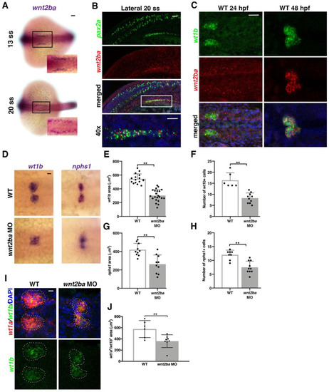
Figure 4. wnt2ba is a podocyte marker and regulator. (A) wnt2ba is expressed in bilateral stripes as early as 13 ss. Scale bar is 30 μm. (B) wnt2ba (red) is expressed within the anteriormost region of the IM, as shown by colocalization with pax2a (green). White box denotes area of colocalization, which is magnified in bottom panel. DAPI (blue) marks nuclei. Scale bar is 15 μm. (C) wnt2ba (red) also colocalized with the podocyte marker wt1b at 24 hpf, though at this time point it was also expressed in the putative neck segment domain. By 48 hpf, the wnt2ba domain was specified to the podocytes. Scale bar is 30 μm. (D–H) Podocyte area and cell number was assessed in wnt2ba morpholino-injected animals and determined to be reduced compared with WT controls. Both wt1b and nphs1 showed a significant decrease in domain area in wnt2ba morphants compared with WT embryos. (I,J) FISH with wt1a and wt1b was performed at 24 hpf. There was a significant area reduction in the podocyte domain seen in wnt2ba morphants compared to WTs. Scale bar is 10 μm. p-values: ** p < 0.001. Scale bar is 30 μm.
|
Expression Data
Expression Detail
Antibody Labeling
Phenotype Data
Phenotype Detail
Acknowledgments
This image is the copyrighted work of the attributed author or publisher, and
ZFIN has permission only to display this image to its users.
Additional permissions should be obtained from the applicable author or publisher of the image.
Full text @ Biomedicines