FIGURE

FIGURE 6

ID
ZDB-FIG-220921-64
Publication
Field et al., 2022 - Involvement of transmembrane protein 184a during angiogenesis in zebrafish embryos
Other Figures
All Figure Page
Back to All Figure Page
FIGURE 6

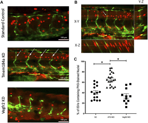
Tmem184a KD Causes an Increase in Cell Proliferation. (A) Example images of PH3 staining in SC, Tmem184a, and Vegfr2 MO knockdown embryos at 48 hpf. White arrows indicate where the PH3 staining co-localizes with the GFP in the vasculature of the ATG MO-treated embryo, highlighted in yellow. Z-stack images were rotated to confirm nuclear PH3 in the EC. Embryos injected with the Tmem184a MO (n = 21) exhibit an increase in the percentage of ISVs that contain an PH3 stained nucleus compared to either SC (n = 21) or Vegfr2 (n = 11) injected embryos. (B) One PH3 nucleus in an ISV is shown in all planes. Cross-hairs intersect at the stained ISV localized nucleus. (C) The graph illustrates the percentage of ISVs per embryo containing an PH3-stained nucleus. p < 0.001.

Expression Data

Expression Detail
Antibody Labeling
Phenotype Data
Fish:
Knockdown Reagents:
Observed In:
Stage: Long-pec

Phenotype Detail
Acknowledgments
This image is the copyrighted work of the attributed author or publisher, and ZFIN has permission only to display this image to its users. Additional permissions should be obtained from the applicable author or publisher of the image. Full text @ Front. Physiol.