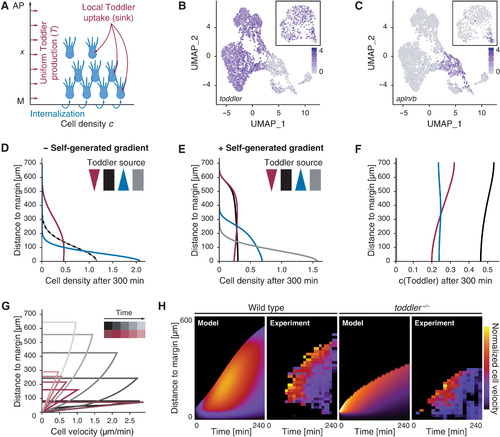
Fig. 4. Computational simulations predict a self-generated Toddler gradient. (A) Schematic representation of the one-dimensional model of mesoderm density and Toddler concentration along the animal-vegetal axis (x = 0 denotes the margin, at which mesodermal cells are added). Toddler (red) is produced uniformly at rate T0 and is degraded locally by mesodermal cells (blue). (B and C) Uniform manifold approximation and projection (UMAP) clustering of single cells at 60% epiboly based on scRNA-seq data (24). Inset depicts UMAP at 30% epiboly. Color code represents expression levels of toddler (B) and aplnrb (C) in individual cells. (D and E) Predicted mesoderm density profiles (arbitrary units) after 300 min without (D) or with (E) Toddler uptake by mesoderm, for different profiles of Toddler production T0(x): graded toward the margin (blue), graded toward the animal pole (red), uniform (black), or no production (gray). (F) Predicted Toddler concentrations (arbitrary units) after 300 min with Toddler uptake by mesoderm. Profiles of Toddler productions T0(x) as described in (D) and (E). (G) Predicted spatiotemporal profiles of mesodermal cell velocities in wild-type (black) and toddler−/− embryos (red). (H) Predicted (model) and experimental (experiment) kymographs of mesodermal cell migration in wild-type (left) and toddler−/− (right) embryos. Experimental data from light sheet microscopy and tracking of drl:GFP-positive cells [N = 7 (wild type) and N = 6 (toddler−/−) embryos; average number of n = 195 cells tracked per embryo]. Color code represents normalized velocity (yellow: high; dark purple: low).
|
