|
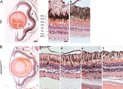
Hematoxylin/eosin histology staining of <italic toggle='yes'>serpine3</italic><sup>cbg18</sup> eyes (14 months) reveals histological differences in comparison to their wild type (WT) siblings (dorsal top, ventral bottom).We show details of representative overview images for one eye of each genotype on the right. In comparison to WT (A), distance between lens and retina of serpine3cbg18 fish is reduced (distance bars). In the serpine3cbg18 eye (B), all retinal layers are present and distinguishable, although they are not as tightly packed and clearly separated as in the WT (c–f). Moreover, we observed displaced pigmented cells that are located mainly in the photoreceptor outer segment and the outer nuclear layer yellow arrows, (c–e). Furthermore, the photoreceptor outer segment and RPE layer are not clearly separated in the serpine3cbg18 retina. Scale bar in the overviews represents 200 µm and in the magnifications 20 µm.
|