
FIGURE
Fig. 5
- ID
- ZDB-FIG-220803-31
- Publication
- Cui et al., 2022 - PARD3 gene variation as candidate cause of nonsyndromic cleft palate only
- Other Figures
- All Figure Page
- Back to All Figure Page
Fig. 5
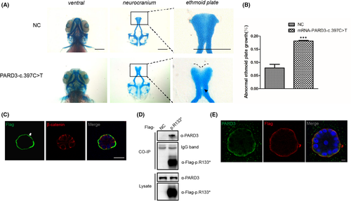
Functional validation of the N-terminal truncating variant c.397C>T identified in a sporadic case with NSCP. (A) Representative images of the control group and zebrafish larvae injected with PARD3-c.397C>T variant mRNA are depicted. Scale bar = 200 μm. (B) Statistical analysis for (A). Bars indicate the means ± SEM. Student’s t test was used to analyse the data. ***p < 0.001. (C) MCF-10A cells stably expressing PARD3-c.397C>T were polarized and formed apical lumens after 10 days of 3D culture in Matrigel. The cysts were stained for Flag-tagged PARD3-c.397C>T (green) and the basolateral membrane marker β-catenin (red). PARD3-c.397C>T localized to the basal compartment rather than the apical region. The arrowhead points to the presence of truncated PARD3 at the basal region. The asterisk indicates the apical area. Scale bar = 25 μm. (D) Endogenous PARD3 interacted with Flag-tagged PARD3-p.R133*. Flag-tagged PARD3-p. R133* was immunoprecipitated from cell lysates of HEK-293T cells stably expressing Flag-tagged PARD3-p. R133*, and the coimmunoprecipitation product was analysed by anti-PARD3 (C-terminal immunogen) and anti-Flag immunoblotting. (E) 3D culture of MCF-10A cells was performed, and MCF-10A cells stably expressing Flag-tagged PARD3-c. 397C>T showed that a substantial proportion of endogenous PARD3 colocalized with PARD3-c.397C>T, which was mainly at the basement membrane. Endogenous PARD3 was analysed with immunofluorescent staining using a C-terminal PARD3 antibody (green), PARD3-c.397C>T was visualized with a Flag antibody (red), and nuclei were stained with DAPI (blue). Scale bar = 7.5 μm
|
Expression Data
Expression Detail
Antibody Labeling
Phenotype Data
Phenotype Detail
Acknowledgments
This image is the copyrighted work of the attributed author or publisher, and
ZFIN has permission only to display this image to its users.
Additional permissions should be obtained from the applicable author or publisher of the image.
Full text @ J. Cell. Mol. Med.