
Fig. 1
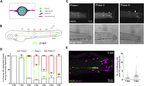
Active cell proliferation and clustering occur during neuromast regeneration upon fin amputation. (A) A cartoon shows different cell types in a neuromast of the lateral line. (B) A graph shows only one side of the fluorescent posterior lateral (green) with L1-8 neuromasts of an Et(HG7L) larva at three days post-fertilization. The tail fin is clipped at the dashed line to remove neuromast L6-8. (C) A new neuromast was regenerated in three distinct phases as examined at the GFP channel under epifluorescent microscopy. Phase I: No notable increase in fluorescent cells was observed in the lateral line between the L5 neuromast (as labeled) and the cut site (dotted line). Phase II: Fluorescent cells were increased and aggregated to form a cluster (arrowhead). Phase III: A new neuromast was formed (arrow). The corresponding bright-field images for each phase are shown below. (D) The percentages of larvae at each phase were calculated on the designated day postamputation (dpa, N = 3, n = 70). (E) Et(HG7L) larvae were fin-amputated, fixed at 1 and 2 dpa, and subjected to BrdU or EdU staining (in magenta) to probe actively proliferating cells or GFP immunohistochemistry (in green) to stain lateral line. Active cell proliferation was observed near the cutting edge at 2 dpa and quantified in a scatter plot on the right. |