
Fig. 4
- ID
- ZDB-FIG-220715-52
- Publication
- Berger et al., 2022 - Mob4-dependent STRIPAK involves the chaperonin TRiC to coordinate myofibril and microtubule network growth
- Other Figures
- All Figure Page
- Back to All Figure Page
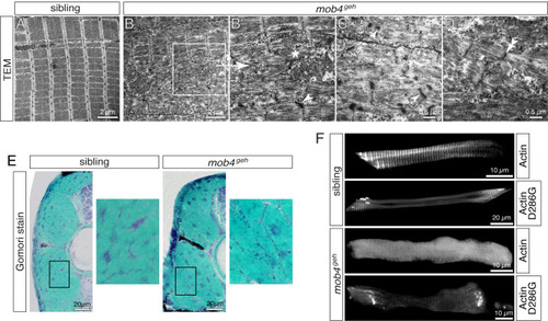
Sarcomere organisation is compromised in mob4geh mutants.
(A) Transmission electron micrograph depicted highly organised and arrayed myofibrils in 3-dpf-old siblings (n = 3). (B) Organised sarcomeres were rarely detected within mob4geh homozygotes (n = 3). (B’) As shown in the magnification of the boxed area, sarcomeres were frequently disorganised and deposits of isolated filaments (asterisk) in addition to electron-dense structures (arrowhead), often associated with Z-disks, were found instead. (C) Electron-dense aggregates of mob4geh homozygotes often showed a lattice structure (arrowhead) and (D) fragmented sarcomeres and widened Z-disks (double-arrowhead) were detected as well. (E) At 3 dpf, Gomori trichrome staining revealed subsarcolemmal dark blue structures within mob4geh homozygotes but not siblings (n = 6 per genotype). Boxed areas are shown in higher magnification. (F) GFP fluorescence of transgenic ACTA1-GFP showed a striated pattern in 3-dpf-old siblings and a uniform pattern in mob4geh homozygotes. Expression of ACTA1D286G-GFP led to rod-shaped structures in siblings and exclusively amorphic aggregates within mob4geh homozygotes (n = 6 per genotype). Scale bar sizes are indicated. |
| Fish: | |
|---|---|
| Observed In: | |
| Stage: | Protruding-mouth |