
Fig. 7
- ID
- ZDB-FIG-220705-59
- Publication
- Lu et al., 2022 - Fish female-biased gene cyp19a1a leads to female antiviral response attenuation between sexes by autophagic degradation of MITA
- Other Figures
- All Figure Page
- Back to All Figure Page
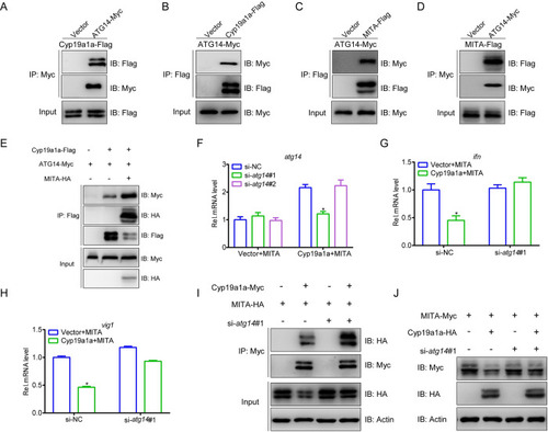
ATG14 is essential for Cyp19a1a mediated MITA autophagic degradation.
(A-D) EPC cells were transfected with the indicated plasmids (5 μg each). After 24 h, cell lysates were IP with anti-Myc or anti-Flag affinity gel. Then the immunoprecipitates and WCLs were analyzed by IB with the anti-Myc and anti-Flag Abs, respectively. (E) EPC cells were transfected with the indicated plasmids (4 μg each). After 24 h, cell lysates were IP with anti-Flag affinity gel. Then the immunoprecipitates and WCLs were analyzed by IB with the anti-Myc, anti-HA, and anti-Flag Abs, respectively. (F) EPC cells were transfected with 100 nM si-atg14#1, si-atg14#2, or si-NC (negative control). At 6 h post-transfection, the cells were transfected with 1.5 μg MITA-Myc plus 1.5 μg Cyp19a1a-Flag or empty vector. At 24 h post-transfection, total RNAs were extracted to examine the transcriptional levels of ATG14. (G and H) EPC cells were transfected with 100 nM si-NC or si-atg14#1. At 24 h post-transfection, cells were co-transfected with 1.5 μg MITA-Myc and 1.5 μg Cyp19a1a-Flag or empty vector for 24 h before qPCR analysis was performed. The relative transcriptional levels were normalized to the transcription of β-actin and represented as fold induction relative to the transcriptional level in control cells, which was set to 1. (I) EPC cells were transfected with 100 nM si-NC or si-atg14#1. After 24 h, the cells were transfected by the indicated plasmids (5 μg each) for 24 h. Cell lysates were IP with anti-Myc affinity gel. Then the immunoprecipitates and WCLs were analyzed by IB with the indicated Abs. (J) EPC cells were transfected with 100 nM si-NC or si-atg14#1. At 24 h post-transfection, cells were co-transfected with 1.5 μg MITA-Myc plus 1.5 μg Cyp19a1a-HA or empty vector for 24 h. The cell lysates were subjected to IB with anti-Myc, anti-HA, and anti-β-actin Abs. |