
Fig. 4
- ID
- ZDB-FIG-220603-61
- Publication
- Biagini et al., 2022 - Zebrafish Melanoma-Derived Interstitial EVs Are Carriers of ncRNAs That Induce Inflammation
- Other Figures
- All Figure Page
- Back to All Figure Page
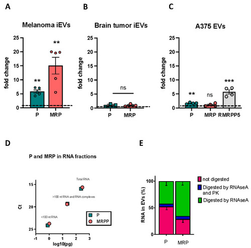
QPCR validation of P and MRP RNAs enrichment in iEVs. (A–C) Expression of P and MRP (and RMRPP5 in human cells/EVs) RNA in melanoma iEVs compared to the expression in melanoma (A), in brain tumour iEVs compared to the expression in tumour cells (B), and in A375 EVs versus A375 cells (C). In all graphs, expression in the cells of origin of the iEVs/EVs is set at 1, and data are normalized to the levels of rnu6.1/RNU6. n > 3 as represented; ** p < 0.01; *** p < 0.001. (D) Graphic representation of the amount of P and MRP found in the lower fraction (fragmented RNA), higher fraction (RNA and RNA–protein complexes), and total RNA of A375 EVs, following UV crosslinking. Values are expressed in pg and are calculated from the number of ct of a reference scale obtained with serial dilutions of P and MRP cDNA templates (see Section 4.16). (E) Percentages of P and MRP RNA lost in RNAseA digested and RNAseA + proteinase K digested in A375 EVs, as indicated. Data are from 3 biological replicates +/− SD. ns: not significant. |