FIGURE

FIGURE 2

ID
ZDB-FIG-220522-2
Publication
Zhang et al., 2022 - Using Light-Sheet Microscopy to Study Spontaneous Activity in the Developing Lateral-Line System
Other Figures
All Figure Page
Back to All Figure Page
FIGURE 2

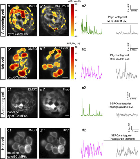
P2yr1 signaling is required for spontaneous activity in supporting cells but not immature hair cells. The spatial patterns of the mean spontaneous calcium activities of the supporting cells (A1–A1′) or hair cells (B1–B1′) in DMSO and after 15 min of treatment with 1 µM MRS2500. Measurements were performed in immature neuromasts at day 3. The ΔF/F GCaMP6s signals were averaged over each 900 s interval (pre- and post-treatment) and then colorized according to the heat map and superimposed onto a baseline image. The corresponding temporal curves of the mean signal magnitude across the whole neuromast in supporting cells (A2) and in hair cells (B2) in DMSO and after 15 min of treatment with 1 µM MRS2500. The cytosolic baseline calcium in the supporting cells (C1–C1′) and hair cells (D1–D1′) in DMSO and after 15 min of treatment with 250 nM Thapsigargin. Measurements were performed in immature neuromasts at day 3. The corresponding temporal curves of the mean signal magnitude across the whole neuromast in supporting cells (C2) and in hair cells (D2) in DMSO and after 15 min of treatment with 250 nM Thapsigargin. Scale bar = 5 μm.

Expression Data

Expression Detail
Antibody Labeling
Phenotype Data

Phenotype Detail
Acknowledgments
This image is the copyrighted work of the attributed author or publisher, and ZFIN has permission only to display this image to its users. Additional permissions should be obtained from the applicable author or publisher of the image. Full text @ Front Cell Dev Biol