
Fig. 4.
- ID
- ZDB-FIG-220422-26
- Publication
- Rutherford et al., 2022 - A zebrafish reporter line reveals immune and neuronal expression of endogenous retrovirus
- Other Figures
- All Figure Page
- Back to All Figure Page
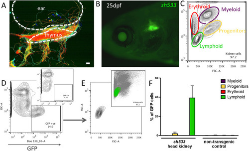
zferv1a is expressed in cells from the lymphoid lineage in adult haematopoietic tissues. (A) Cell tracking reveals dynamic behaviour of entry and exit of the thymus by GFP-positive cells in Tg(zferv1a:GFP)sh533. Whole-stack analysis of cell movement using TrackMate in Fiji. Scale bar: 10 µm. (B) Expression of Tg(zferv1a:GFP)sh533 in a 25 dpf zebrafish highlighting the triangular shaped thymus. (C) Forward scatter (FSC)/side scatter (SSC) plot of a whole kidney marrow separating different haematopoietic lineages by size and granularity. (D) SSC and GFP plot from a single-cell suspension of a whole kidney marrow from an adult Tg(zferv1a:GFP) with gate selecting GFP-positive cells. Inset shows the distribution of cells from a non-transgenic adult whole kidney marrow. (E) FSC/SSC plot of selected GFP-positive cells from D. Inset shows the location of the GFP-positive cells in the haematopoietic lineage FSC/SSC plot. (F) Quantification of GFP-positive cells in myeloid, erythroid, lymphoid lineages and progenitors from dissected sh533 and non-transgenic control adult head kidneys (n=3). |