
Fig. 1
- ID
- ZDB-FIG-220203-70
- Publication
- Tajer et al., 2021 - BMP heterodimers signal via distinct type I receptor class functions
- Other Figures
- All Figure Page
- Back to All Figure Page
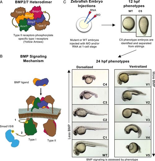
BMP heterodimer signaling and assays in the zebrafish embryo. (A) A BMP heterodimer contains four distinct receptor binding sites for two type I and two type II receptors. One type I receptor site resembles the BMP2 homodimer type I receptor binding site, predicted to bind Bmpr1. The other type I receptor binding site resembles the BMP7 homodimer type I receptor binding site, predicted to bind Acvr1. Yellow arrows indicate the type II-type I receptor interactions. (B) The BMP signaling mechanism: Type II receptors phosphorylate Type I receptors (1), which in turn phosphorylate and activate the transcription factor Smad1/5/8 (2). (C) Experimental schematic. Eggs are injected at the one-cell stage. Embryos are screened, photographed, and C5 phenotype embryos separated at 12 hpf. After 24 hpf, strongly dorsalized C5 embryos have died, and all other phenotypes are photographed and classified. |