
- Title
-
BMP heterodimers signal via distinct type I receptor class functions
- Authors
- Tajer, B., Dutko, J.A., Little, S.C., Mullins, M.C.
- Source
- Full text @ Proc. Natl. Acad. Sci. USA
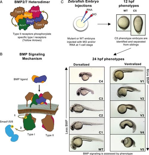
BMP heterodimer signaling and assays in the zebrafish embryo. (A) A BMP heterodimer contains four distinct receptor binding sites for two type I and two type II receptors. One type I receptor site resembles the BMP2 homodimer type I receptor binding site, predicted to bind Bmpr1. The other type I receptor binding site resembles the BMP7 homodimer type I receptor binding site, predicted to bind Acvr1. Yellow arrows indicate the type II-type I receptor interactions. (B) The BMP signaling mechanism: Type II receptors phosphorylate Type I receptors (1), which in turn phosphorylate and activate the transcription factor Smad1/5/8 (2). (C) Experimental schematic. Eggs are injected at the one-cell stage. Embryos are screened, photographed, and C5 phenotype embryos separated at 12 hpf. After 24 hpf, strongly dorsalized C5 embryos have died, and all other phenotypes are photographed and classified. |

BMP homodimers fail to rescue embryos lacking BMP antagonists and heterodimers: In bubble plots, circle area reflects the percent embryos of a given phenotype (Left), fill color reflects the MO condition, and line color reflects the RNA injection condition. Ns are in brackets above each column. Injection conditions and genotypes are labeled below each column. Raw phenotype scores are shown in Dataset S1. MO concentrations and combinations are listed in SI Appendix, Table S4. Hypothesis 1: BMP heterodimers prevail because antagonists preferentially bind homodimers (A) and, if true, then BMP homodimers should signal in the absence of antagonists and heterodimers (B). (C) Bmp2 homodimers cannot signal at endogenous expression levels, even without CNF. Column 1: 1/4 of the offspring of a bmp2b+/− incross are C5 bmp2b−/−. Column 2: 15 pg FLAG-bmp2b RNA injection rescues most C5 mutant embryos and ventralizes nonmutants. Other columns: bmp2b homodimers fail to rescue bmp7a mutant embryos with or without CNF and additional bmp2b RNA. (D–H) Column numbers below images refer to the experimental condition of columns in C. The otx2b expression can be seen in the following: WT embryos (D), bmp7a mutant embryos (E), bmp7a mutants with additional bmp2b RNA (F), CNF MO-injected bmp7a mutants (G), and bmp7a mutants injected with CNF MO and bmp2b RNA (H). (I) Bmp7 homodimers cannot signal at endogenous concentrations, even without CNF. Column 1: bmp7a−/− embryos are C5 dorsalized. Column 2: 40 pg bmp7a RNA rescues DV patterning in bmp7a mutants. Other columns: Bmp7 homodimers fail to rescue bmp2b mutants with or without CNF and additional bmp7a RNA. |

Overexpressed Bmp2 homodimers require both Bmpr1 and Acvr1 to signal: The HA-bmp2b RNA injected is 5- to 40-fold (5 to 10 pg) above the rescuing concentrations (0.25 to 1 pg) shown in SI Appendix, Fig. S4. Raw phenotype scores are shown in Dataset S2. MO concentrations and combinations are listed in SI Appendix, Table S4. (A) Overexpressed Bmp2 homodimers require Bmpr1 to signal. The bmp2b overexpression ventralizes bmp7−/− embryos (columns 1 and 2). The bmpr1a MO knockdown abates signaling caused by bmp2b overexpression (column 3). Efficacy of the bmpr1a MO mix is affirmed in WT embryos (columns 4 and 5). (B) In one model, the BMP2 homodimer forms a Bmpr1–Bmpr1 complex. (C) Overexpressed Bmp2 homodimers require Acvr1 to signal. Overexpressed bmp2b RNA ventralizes bmp7−/− embryos (columns 1 and 2). The acvr1l MO knockdown abates signaling caused by bmp2b overexpression (column 3). Efficacy of the acvr1l MO is affirmed in WT embryos too (columns 4 and 5). (D) In another model, the BMP2 homodimer binds to Acvr1 and Bmpr1. (E) Bmpr1–Acvr1 heteromeric complex is necessary for BMP2 homodimer signaling in the zebrafish. |
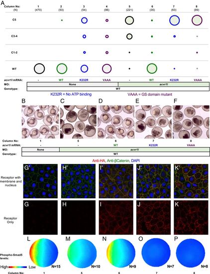
Acvr1l kinase function is essential for BMP signaling. (A) Kinase-dead Acvr1l cannot signal and restore DV patterning. WT embryos injected with acvr1l-HA RNA (100 pg), kinase-dead acvr1l-K232R-HA RNA (60 pg), and GS-domain mutant acvr1VAAA-HA RNA (100 pg), with or without acvr1l MO. MO concentrations and combinations are listed in SI Appendix, Table S4. Raw phenotype scores are in Dataset S3. (B–F) The acvr1l MO-injected embryos at ∼24 hpf. Injection conditions and corresponding columns in A are labeled below the images. Uninjected embryos are WT (B) Acvr1l-deficient C5 embryos lyse by 24 hpf (C). (D) WT acvr1l-HA RNA fully rescues Acvr1l-deficient embryos. (E and F) Acvr1l-deficient embryos injected with (E) acvr1l-K232R-HA RNA or (F) acvr1l-VAAA-HA RNA remain C5 dorsalized and lyse by 24 hpf. (G–K) Immunostaining against HA-tagged Acvr1l receptor with injection conditions labeled above the images. Embryos ≥3 were imaged for each condition. G’–K’ are the same images as G–K but showing DAPI-stained nuclei (blue) and membrane-localized β-catenin (green). (L–P) Quantified and averaged phospho-Smad5 immunostained embryos from various Acvr1l injection conditions. Corresponding columns in A are labeled on the Bottom. (L) Uninjected embryos display a WT phospho-Smad5 gradient. (M) Acvr1l-deficient embryos display reduced phospho-Smad5. (N) WT acvr1l RNA restores phospho-Smad5 signaling in Acvr1l-deficient embryos, while acvr1l-K232R RNA (O) and acvr1l-VAAA RNA (P) do not. |

Kinase-dead Bmpr1 can restore Bmp signaling in Bmpr1-deficient embryos: (A and B) The bubble-plot fill color reflects genotype condition. Raw phenotype scores are in Dataset S4. (A) Kinase-dead Bmpr1 can rescue bmpr1aa−/−; bmpr1ab−/− double mutant embryos. Column 1: uninjected bmpr1aa+/+; bmpr1ab−/− and bmpr1aa+/−; bmpr1ab−/− embryos are phenotypically WT. Columns 2-4: bmpr1aa+/−; bmpr1ab−/− embryos injected with WT bmpr1aa RNA (40 to 80 pg) (column 2), kinase-dead bmpr1aa-K256R RNA (20 to 80 pg) (column 3), or GS-domain mutant bmpr1aa-VVAAA RNA (80 pg) (column 4). Column 5: bmpr1aa−/−; bmpr1ab−/− double mutants are C4 dorsalized. Columns 6 to 8: bmpr1aa−/−; bmpr1ab−/− double mutants injected with WT bmpr1aa RNA (column 6), bmpr1aa-K256R RNA (column 7), or bmpr1aa-VAAA RNA (column 8). (B–F) Representative-rescued embryos from bmpr1 genotypes and injection conditions in the following: a bmpr1aa+/−; bmpr1ab−/− embryo (A and B), bmpr1aa−/−; bmpr1ab−/− double mutants (C–F), uninjected (C), injected with WT bmpr1aa RNA (D), injected with bmpr1aa-K256R RNA (E), and injected with bmpr1aa-VAAA RNA (F). (G) Kinase-dead Bmpr1 rescues the simultaneous depletion of all four zebrafish Bmpr1 receptors. Experimental conditions in all columns are as in A, except that all embryos are additionally injected with a combination of bmpr1ba and bmpr1bb MO. MO concentrations and combinations are listed in SI Appendix, Table S4. H–L is the same as B–F but with additional bmpr1ba and bmpr1bb MO injection. |