
FIGURE 2
- ID
- ZDB-FIG-220131-118
- Publication
- Hopfenmüller et al., 2022 - The Wilms Tumor Gene wt1a Contributes to Blood-Cerebrospinal Fluid Barrier Function in Zebrafish
- Other Figures
- All Figure Page
- Back to All Figure Page
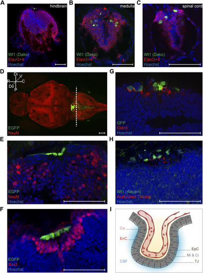
Characterization of Wt1+ cells of the zebrafish larval CNS. (A-C) Transverse sections through the hindbrain, the caudal medulla and spinal cord of 4.25 days old Tg(wt1a:EGFP) zebrafish larvae were stained with antibodies against Wt1 (Dako) and Elavl3+4. The images show optical sections of one focus plane. Orientation: dorsal: up. Scale bar: 50 µm. (D) Whole zebrafish larvae (4.5 dpf) of the Tg(wt1a:EGFP) line were used for detection of the EGFP signal of Wt1 positive cells and IHC against NeuN. The image is displayed as extended depth of focus projection. The fluorescence signal for EGFP does not overlap with NeuN staining. The dotted line indicates the cutting position of the following transverse sections. Orientation: rostral: left; dorsal view. Abbreviations: distal (Di); proximal (P); rostral (R); caudal (C); dorsal (Do); ventral (V) Scale bar: 50 µm. (E) Transverse section through the hindbrain of 4.25 days old Tg(wt1a:EGFP) zebrafish larvae were used for detection of the EGFP signal of Wt1 positive cells and IHC against NeuN. (F) Transverse sections through the hindbrain of 4.25 days old Tg(wt1a:EGFP) zebrafish larvae were used for detection of the EGFP signal of Wt1 positive cells and IHC against Sox2. (G) Transverse sections through the hindbrain of 4.25 days old Tg(wt1a:EGFP) zebrafish larvae were used for IHC against GFP and Cldn5. (H) Transverse sections through the hindbrain of 4.25 days old Tg(wt1a:EGFP) zebrafish larvae were used for IHC against Wt1 (Abcam) and acetylated Tubulin. (E–H) The images show optical sections of one focus plane. Orientation: dorsal: up. Scale bar: 50 µm. (I) Schematic illustration of the ependymal cells of the fourth brain ventricle, which are connected via tight junctions and show microvilli and cilia at their apical surface. The image was created with BioRender.com. Abbreviations: capillary (Ca); endothelial cell (EnC); cerebrospinal fluid (CSF); ependymal cells (EpC); microvilli (Mi); Cilia (Ci); tight junction (TJ). |