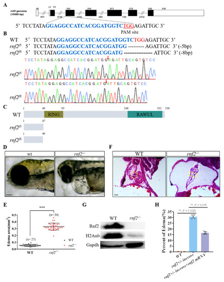
Rnf2-null zebrafish mutant displayed severe cardiac defects. (A) The target site (sequence highlighted in blue) is located in exon 3. The PAM site is underlined and highlighted in red; (B) DNA sequencing identified that two mutant alleles carried a 5 bp (rnf2f5) and 8 bp (rnf2f8) deletion respectively. The deletion sites are indicated by red arrows; (C) Schematic diagram of the wild-type and mutant Rnf2 proteins. Zebrafish wild-type Rnf2 contained an N-terminal Ring-finger domain (yellow) and a C-terminal RAWUL domain. The two mutant proteins were truncated before the RING-finger domain; (D) The rnf2−/− larvae displayed severe pericardia edema. The hearts are indicated by white arrows, the stringy heart in rnf2−/− is depicted by white dotted lines, scale bar: 0.2 mm; (E) Scatter plot showing the sectional area of edema in wild-type (black) and rnf2−/− (red). (F) Histological sections of 4 dpf heart, boxes in yellow indicate the AVC and valves, white boxes indicate the bulbous arteriosus, scale bar: 50 μm, n(WT) = 2, n (rnf2−/−) = 3; (G) Western blot verified the deletion of Rnf2 protein. Gapdh was set as internal reference; (H) Bar graph showing the percentage of embryos with edema. **, p < 0.01; ***, p < 0.001; n, sample number.
|
