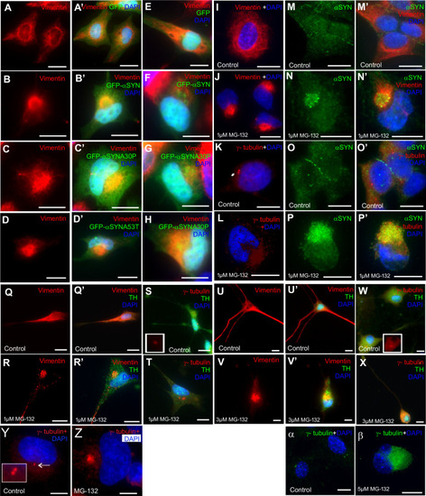
Aggresomes can be formed in cells by overexpression of alpha synuclein or MG132 treatment. (A,A′) Vimentin (red) in control HeLa cells forms a fibrous network around the nuclei (DAPI, blue), which is unaffected by the expression of the GFP. (B,B′) When expressing GFP- α-syn, vimentin-positive aggresomes appear in cells, juxtaposed to the nucleus. Similar results are obtained with GFP-α-synA30P (C,C′) and GFP-α-synA53T mutants (D,D′) and in SH-SY5Y cells treated in the same way (E–H). (I–L) Aggresomes can also be induced by treatment with MG132. (I,J) The vimentin distribution changes from a filamentous pattern around the nuclei to caging the aggresome. (K,L) γ-tubulin (red) staining the centrosomes as two punctae in control cells (K). Following MG132 treatment, γ-tubulin forms a condensed structure around the aggresome (L). (M–P) The expression pattern of endogenous α-syn was also investigated to determine whether this protein co-localises within the aggresome. (M,M′) In control cells, endogenous α-syn (green) staining was widespread and diffuse within the cytoplasm with vimentin (red) forming a filamentous network. (N,N′) Following MG132 treatment, α-syn aggregates were observed and co-localised with vimentin staining within the aggresomes. (O,O′) In control cells, γ-tubulin (red) was observed as two punctae with α-syn diffuse within the cytoplasm. (P,P′) γ-tubulin staining (red) also co-localises with endogenous α-syn in the aggresome when treated with MG132. (Q,Q′) Differentiated SH-SY5Y cells were mock-treated and vimentin (red) staining was observed surrounding the nuclei as well as along the axon. (R,R′) In cells treated with MG132 (1 μM for 18 h) vimentin staining changed to a compact structure near the nucleus, indicative of aggresomes. (S) In mock-treated cells, γ-tubulin formed two punctae next to the nucleus. (T) In MG132 treated cells, aggresomes were detected by γ-tubulin staining. Differentiated SH-SY5Y cells are TH positive (green). (U,U′) In rat basal ganglion neurons, vimentin staining (red) is abundant around the nuclei and along the axon. (V,V′) When treated with MG132, vimentin localises to the aggresome. (W) In rat basal ganglion neurons, the γ-tubulin is observed at two punctae close to the nucleus. (X) Upon MG132 treatment, the γ-tubulin staining now forms a larger structure next to the nucleus. (Y) In untreated RPE1-hTERT cells, γ-tubulin stains the centrosome. (Z) Upon MG132 treatment it stains the aggresome. (α,β) Similar results are obtained with MEFs. Scale bars: 10 μM. DNA/nuclei stained with DAPI (blue) where indicated.
|
