|
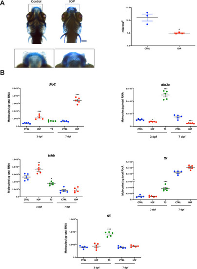
Effects of IOP treatment on thyroid status.(A) Effect of IOP on Meckel´s cartilage morphology. Left: Representative image of Alcian blue staining of 7 dpf control zebrafish (left) and IOP-treated (right) larvae. Scale bar 100 μm. Right: Graph depicting the quantification of Meckel’s cartilage (n = 3 larvae per group). The difference is best visible in the top middle where the stained area is thinner and more faint in the IOP animal. Scale bar 100 μm. Statistical analysis was performed with a Student’s t test, *p<0.01 (B) Changes in expression of TH-regulated genes in zebrafish larvae exposed to IOP. mRNA quantification of dio2, dio3, tshb, ttr and gh in controls and larvae exposed to IOP 0.5 μM or T3 10 nM for 3 dpf and to IOP 0.5 μM for 7 dpf. Data are represented as individual values in vertical graphs (n = 5 pools from independent experiments of 50–60 larvae per pool and condition). Graphs are showing individual values and mean ± SEM. Statistical analysis was performed with one-way ANOVA coupled with Tukey’s multiple comparison test with respect to the corresponding control groups. Significant differences are indicated as *p <0.05, **p <0.01, and ***p <0.001.
|