FIGURE

FIGURE 2

ID
ZDB-FIG-210718-33
Publication
Hsiao et al., 2021 - The Incoherent Fluctuation of Folate Pools and Differential Regulation of Folate Enzymes Prioritize Nucleotide Supply in the Zebrafish Model Displaying Folate Deficiency-Induced Microphthalmia and Visual Defects
Other Figures
All Figure Page
Back to All Figure Page
FIGURE 2

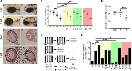
Morphological and functional characterization on the eyes of FD larvae. (A,B) Control and FD larvae at indicated stages with/without folate supplementation were imaged laterally for the head region and quantified for the eye area (dotted circle) with Image J (An on-line software for scientific image analysis.). The larvae in folate supplemented groups were exposed to the indicated folate adducts at 25 hpf and until observation. (C) The diameter of lens was estimated from the images of H&E stained cryo-sections prepared from the larvae at 5 dpf. (D) The experimental flow of optomotor response analysis (OMR). (E) Control and FD larvae with/without folate supplementation were analyzed for OMR at 7 dpf individually and quantified. WT, wild-type; C (control), heat-shocked wild-type larvae; M, FD transgenic larvae with mild FD. S, FD transgenic larvae with severe FD. L, length of lens; ON, optic nerve; RPE, retinal pigmented epithelium; IS/OS, inner segment/outer segment junction; ONL, outer nuclear layer; OPL, outer plexiform layer; INL, inner nuclear layer; IPL, inner plexiform layer; GCL, ganglion cell layer; FA, folic acid (1mM); 5-CHO THF, 5-formyltetrahydrofolate (0.5 mM); 5-CH3 THF, 5-methyltetrahydrofolate (1 mM). Statistical data are shown in mean ± SEM. ***p-value < 0.001; **p-value < 0.01; *p-value < 0.05.

Expression Data

Expression Detail
Antibody Labeling
Phenotype Data
Fish:
Conditions:
Observed In:
Stage Range: Protruding-mouth to Day 5

Phenotype Detail
Acknowledgments
This image is the copyrighted work of the attributed author or publisher, and ZFIN has permission only to display this image to its users. Additional permissions should be obtained from the applicable author or publisher of the image. Full text @ Front Cell Dev Biol