FIGURE

Figure 8

ID
ZDB-FIG-210628-53
Publication
Ishii et al., 2021 - Correlative microscopy and block-face imaging (CoMBI) method for both paraffin-embedded and frozen specimens
Other Figures
All Figure Page
Back to All Figure Page
Figure 8

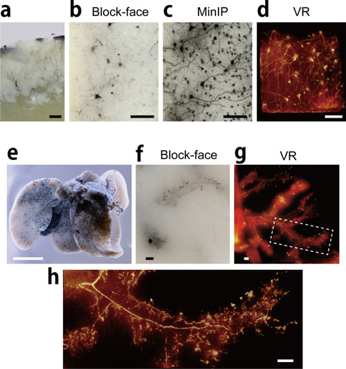
Fine 3D imaging at cellular level. (a) Mouse cerebral cortex was stained by Golgi silver impregnation method. (b) Block-face image of the stained cortex. (c, d) The minimum intensity projection (MinIP) image and volume-rendered image were generated from 201 block-face images with voxel size 0.9 × 0.9 × 1 µm. (e) Mouse liver were injected with black ink via bile duct. (f) A piece of the liver were imaged using CoMBI-S, and a block-face image is shown. (g, h) 3D volume-rendered images using 401 block-face images with voxel size of 4 × 4 × 1 µm (g) or 1 × 1 × 1 µm (h) shows the biliary tree. Bars: 100 µm (ad, fh), 5 mm (e).

Expression Data

Expression Detail
Antibody Labeling
Phenotype Data

Phenotype Detail
Acknowledgments
This image is the copyrighted work of the attributed author or publisher, and ZFIN has permission only to display this image to its users. Additional permissions should be obtained from the applicable author or publisher of the image. Full text @ Sci. Rep.