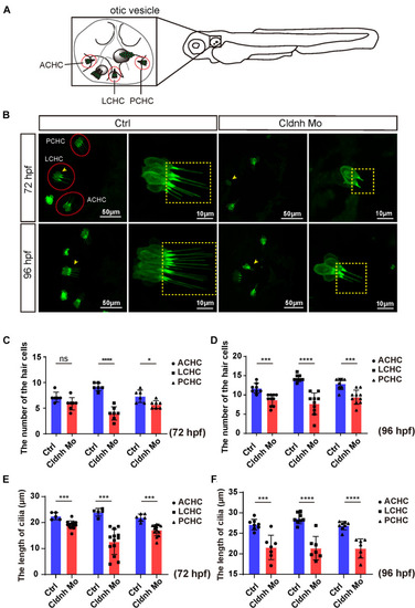
Claudin h deficiency suppressed cristae hair cells development. (A) The schematic for three different cristae hair cells in the otic vesicle. ACHC, anterior cristae hair cells; LCHC, lateral cristae hair cells; PCHC, posterior cristae hair cells. (B) Confocal imaging analysis of cristae hair cells in the otic vesicle of control and claudin h deficiency zebrafish at 72 and 96 hpf. The red dotted circle line marked the three different cristae hair cell clusters and magnified lateral cristae hair cell clusters (yellow arrow head) was shown in right and the yellow dotted square line marked the cilia of cristae hair cells. (C,D) The statistical analysis of the numbers of different cristae hair cells in the control and claudin h morphants at 72 and 96 hpf. (E,F) The statistical analysis of the cilia lengths of different cristae hair cells in the control and claudin h morphants at 72 and 96 hpf. Values with *, ***, and ****above the bars are significantly different (P < 0.05, P < 0.001, and P < 0.0001, respectively).
|
