|
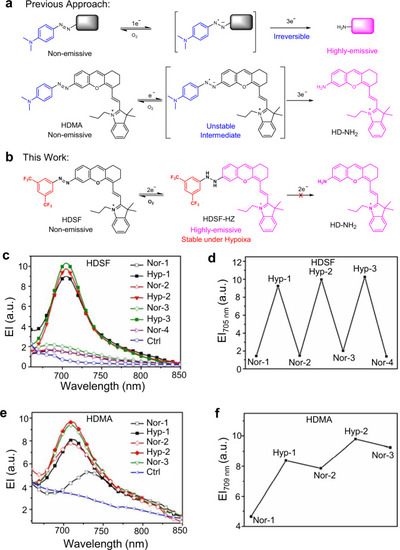
Reversible imaging mechanism and behavior of HDSF.a Normal azobenzene-derived fluorescent probes for hypoxia and their reductive decomposition of N–N bond by the hypoxic microenvironment in living systems. b The reversible hypoxia sensing mechanism of probe HDSF. The square and fluorophore in black represent the quenched-fluorophore, and those in bright magenta represent the emitting-fluorophore. c Fluorescent spectra of 20 μM HDSF in PBS buffer (0.1 M, pH 7.4, 2% DMSO, v/v) containing rat liver microsomes (RLM, 250 μg mL−1) and NADPH (100 μM) recorded in normoxia-hypoxia cycles. d Fluorescence intensity of HDSF at 705 nm detected in (c). e Fluorescent spectra of 20 μM HDMA in PBS buffer (0.1 M, pH 7.4, 2% DMSO, v/v) containing RLM (250 μg mL−1) and NADPH (100 μM) recorded in normoxia-hypoxia cycles. f Fluorescence intensity of HDMA at 709 nm detected in (e). Ctrl: PBS buffer with RLM and NADPH. λex, 650 nm.
|