
Fig. 2
- ID
- ZDB-FIG-210517-11
- Publication
- Schoels et al., 2021 - Single-cell mRNA profiling reveals changes in solute carrier expression and suggests a metabolic switch during zebrafish pronephros development
- Other Figures
- All Figure Page
- Back to All Figure Page
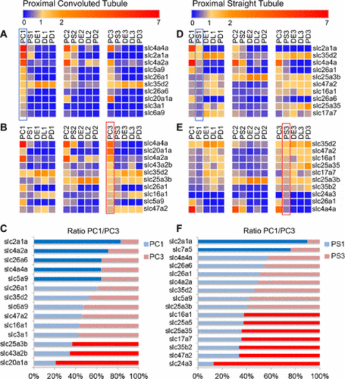
Expression of solute carriers (SLCs) in the proximal convoluted and straight tubule of the developing zebrafish pronephros. A: ten most abundant SLCs in the proximal convoluted tubule at 1 day postfertilization (dpf) (PC1; outlined with blue) compared with the relative expression of these top 10 SLCs in the proximal straight tubule (PS1), distal early tubule (DE1), distal late tubule (DL1), and pronephric duct (PD1) as well as their expression at 2 dpf (PC2, PS2, DE2, DL2, and PD2, respectively) and 3 dpf (PC3, PS3, DE3, DL3, and PD3, respectively). B: 10 most abundant SLCs in PC3 (outlined with red) compared with the relative expression of these top 10 SLCs in PS3, DE3, DL3, and PD3 as well as their expression at 1 dpf and 2 dpf. C: relative change in expression between 1 dpf and 3 dpf for the top 10 candidates identified in the proximal convoluted tubule at 1 dpf and 3 dpf. Blue labeled bars indicate ≥60% abundance at 1 dpf; red labeled bars indicate ≥60% abundance at 3 dpf. The light blue and red bars correspond to <60% abundance. D: 10 most abundant SLCs in PS1 (outlined with blue) compared with the relative expression of these top 10 SLCs in PC1, DE1, DL1, and PD1 as well as their expression at 2 dpf and 3 dpf. E: 10 most abundant SLCs in PS3 (outlined with red) compared with the relative expression of these top 10 SLCs in PC3, DE3, DL3, and PD3 as well as their expression at 1 dpf and 2 dpf. F: relative change in expression between 1 dpf and 3 dpf for the top 10 candidates identified in the proximal straight tubule at 1 dpf and 3 dpf. Blue labeled bars indicate ≥60% abundance at 1 dpf; red labeled bars indicate ≥60% abundance at 3 dpf. The light blue and red bars correspond to <60% abundance. PC3; pronephric cells. |