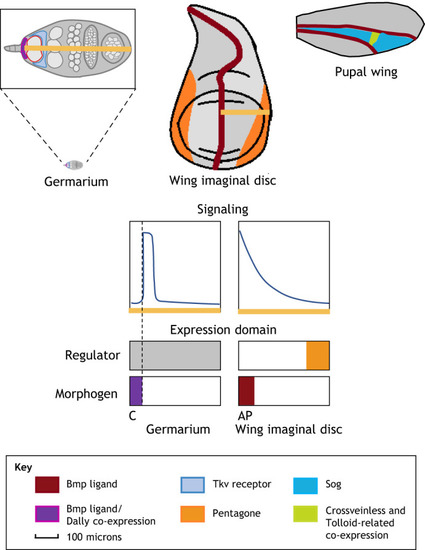
Signaling gradient profiles and expression domains of BMP-patterned organs of different scales.Drosophila germarium (top left): BMP/Dpp and Dally are co-expressed in cap cells (purple). Type IV Collagen Vkg (not pictured) is expressed throughout the GSC niche. Tkv is highly expressed in somatic escort cells. GSC cells are outlined in red (image modified from Sun et al., 2010). Drosophila third instar imaginal wing disc (top middle): BMP/Dpp is expressed in a narrow stripe at the AP boundary. Pentagone is expressed at the periphery. Crossvein formation in Drosophila pupal wing disc (top right): BMP/Dpp is expressed in longitudinal veins, Sog is expressed throughout the pupal wing. Crossveinless and Tolloid-related are expressed in the future posterior cross vein location where they can act to promote BMP/Dpp signaling by liberating ligand from Sog-Dpp complexes. Lower schematics show qualitative graphs of BMP signaling gradients and the expression domains for the morphogen and negative regulator. Germarium regulator depicted as a gray box to reflect ubiquitous presence of multiple regulators including Type IV Collagen Vkg. Signaling and expression domain graphs not shown for pupal wing disc as active transport does not take place over a single axis. AP, anterior posterior boundary; C, cap cells.
|
